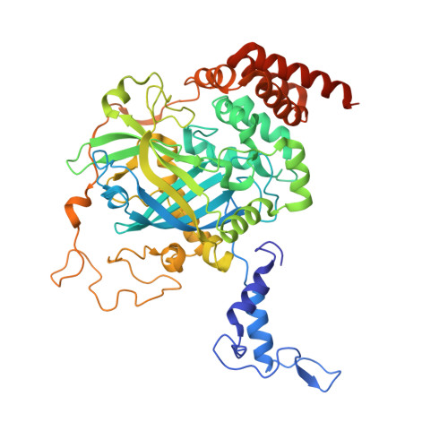
Interaction of human erythrocyte catalase with air-water interface in cryoEM.
Chen, S., Li, J., Vinothkumar, K.R., Henderson, R.(2022) Microscopy (Oxf) 71: i51-i59
- PubMed: 35275189 Search on PubMedSearch on PubMed Central
- DOI: https://doi.org/10.1093/jmicro/dfab037
- Primary Citation Related Structures:
7P8W - PubMed Abstract:
One of the key goals in single-particle cryo-microscopy is to obtain a uniform distribution of particle orientations, so that the three-dimensional structure has isotropic resolution in Fourier space. A common problem arises from the interaction of protein molecules with the air-water interface that exists on both surfaces of the thin film of liquid that is formed prior to plunge-freezing into liquid ethane. Some proteins and other macromolecular complexes are disrupted by interaction with the air-water interface. Other proteins or macromolecules either become concentrated through their interaction with the interface or are excluded because they bind strongly to some other part of the grid or the filter paper used in blotting. In this paper, the interaction of human erythrocyte catalase with the air-water interface is investigated and minimized by the addition of certain detergents. Detergents can form an amphipathic monolayer at the air-water interface that creates a barrier and leaves the molecules free to adopt a variety of orientations, thus facilitating the 3D structure determination. These results suggest that further characterization and development of detergents for cryo-microscopy plunge-freezing would be useful.
- MRC Laboratory of Molecular Biology, Francis Crick Avenue, Cambridge CB2 0QH, UK.
Organizational Affiliation: