7P8V

The structure of E. coli MutL bound to a 3' resected DNA end


Experimental Data Snapshot

  • Method: ELECTRON MICROSCOPY
  • Resolution: 3.60 Å
  • Aggregation State: PARTICLE 
  • Reconstruction Method: SINGLE PARTICLE 

wwPDB Validation   3D Report Full Report

Currently 7P8V does not have a validation slider image.


This is version 1.1 of the entry. See complete history


Literature

MutL binds to 3' resected DNA ends and blocks DNA polymerase access.

Borsellini, A.Lebbink, J.H.G.Lamers, M.H.

(2022) Nucleic Acids Res 50: 6224-6234

  • DOI: https://doi.org/10.1093/nar/gkac432
  • Primary Citation of Related Structures:  
    7P8V

  • PubMed Abstract: 

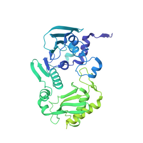
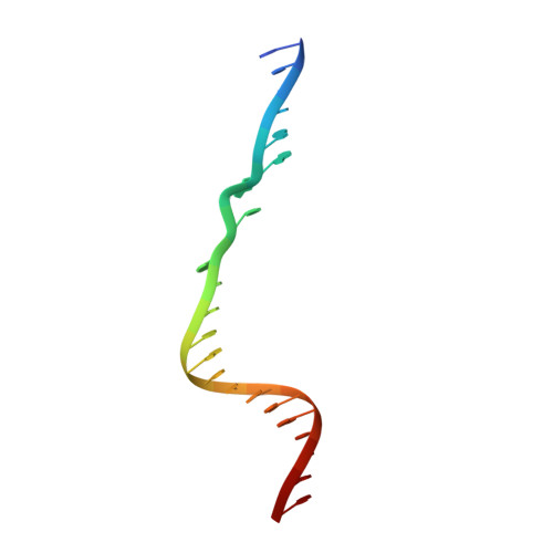
    DNA mismatch repair removes mis-incorporated bases after DNA replication and reduces the error rate a 100-1000-fold. After recognition of a mismatch, a large section of up to a thousand nucleotides is removed from the daughter strand followed by re-synthesis. How these opposite activities are coordinated is poorly understood. Here we show that the Escherichia coli MutL protein binds to the 3' end of the resected strand and blocks access of Pol I and Pol III. The cryo-EM structure of an 85-kDa MutL-DNA complex, determined to 3.7 Å resolution, reveals a unique DNA binding mode that positions MutL at the 3' end of a primer-template, but not at a 5' resected DNA end or a blunt DNA end. Hence, our work reveals a novel role for MutL in the final stages of mismatch repair by preventing premature DNA synthesis during removal of the mismatched strand.


  • Organizational Affiliation

    Department of Cell and Chemical Biology, Leiden University Medical Center, Leiden, The Netherlands.


Macromolecules

Find similar proteins by:  (by identity cutoff)  |  3D Structure
Entity ID: 1
MoleculeChains Sequence LengthOrganismDetailsImage
DNA mismatch repair protein MutL
A, B
615Escherichia coli K-12Mutation(s): 0 
Gene Names: mutLb4170JW4128
UniProt
Find proteins for P23367 (Escherichia coli (strain K12))
Explore P23367 
Go to UniProtKB:  P23367
Entity Groups  
Sequence Clusters30% Identity50% Identity70% Identity90% Identity95% Identity100% Identity
UniProt GroupP23367
Sequence Annotations
Expand
  • Reference Sequence

Find similar nucleic acids by:  Sequence   |   3D Structure  

Entity ID: 2
MoleculeChains LengthOrganismImage
Template strand22DNA molecule
Sequence Annotations
Expand
  • Reference Sequence

Find similar nucleic acids by:  Sequence   |   3D Structure  

Entity ID: 3
MoleculeChains LengthOrganismImage
Primer strand13DNA molecule
Sequence Annotations
Expand
  • Reference Sequence
Experimental Data & Validation

Experimental Data

  • Method: ELECTRON MICROSCOPY
  • Resolution: 3.60 Å
  • Aggregation State: PARTICLE 
  • Reconstruction Method: SINGLE PARTICLE 
EM Software:
TaskSoftware PackageVersion
RECONSTRUCTIONRELION3.1

Structure Validation

View Full Validation Report

Currently 7P8V does not have a validation slider image.


Entry History & Funding Information

Deposition Data


Funding OrganizationLocationGrant Number
Marie Sklodowska-Curie Actions, FragNET ITNEuropean Union--

Revision History  (Full details and data files)

  • Version 1.0: 2022-06-29
    Type: Initial release
  • Version 1.1: 2022-08-31
    Changes: Database references