Spectroscopic/Computational Characterization and the X-ray Structure of the Adduct of the V IV O-Picolinato Complex with RNase A.
Ferraro, G., Demitri, N., Vitale, L., Sciortino, G., Sanna, D., Ugone, V., Garribba, E., Merlino, A.(2021) Inorg Chem 60: 19098-19109
- PubMed: 34847328 Search on PubMedSearch on PubMed Central
- DOI: https://doi.org/10.1021/acs.inorgchem.1c02912
- Primary Citation Related Structures:
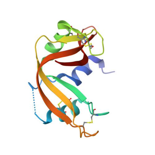
7P8R - PubMed Abstract:
The structure, stability, and enzymatic activity of the adduct formed upon the reaction of the V-picolinato (pic) complex [V IV O(pic) 2 (H 2 O)], with an octahedral geometry and the water ligand in cis to the V═O group, with the bovine pancreatic ribonuclease (RNase A) were studied. While electrospray ionization-mass spectrometry, circular dichroism, and ultraviolet-visible absorption spectroscopy substantiate the interaction between the metal moiety and RNase A, electron paramagnetic resonance (EPR) allows us to determine that a carboxylate group, stemming from Asp or Glu residues, and imidazole nitrogen from His residues are involved in the V binding at acidic and physiological pH, respectively. Crystallographic data demonstrate that the V IV O(pic) 2 moiety coordinates the side chain of Glu111 of RNase A, by substituting the equatorial water molecule at acidic pH. Computational methods confirm that Glu111 is the most affine residue and interacts favorably with the OC -6-23-Δ enantiomer establishing an extended network of hydrogen bonds and van der Waals stabilizations. By increasing the pH around neutrality, with the deprotonation of histidine side chains, the binding of the V complex to His105 and His119 could occur, with that to His105 which should be preferred when compared to that to the catalytically important His119. The binding of the V compound affects the enzymatic activity of RNase A, but it does not alter its overall structure and stability.
- Department of Chemical Sciences, University of Naples Federico II, I-80126 Napoli, Italy.
Organizational Affiliation: