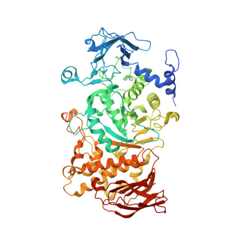
The Candida glabrata glycogen branching enzyme structure reveals unique features of branching enzymes of the Saccharomycetaceae phylum.
Conchou, L., Martin, J., Goncalves, I.R., Galisson, F., Violot, S., Guilliere, F., Aghajari, N., Ballut, L.(2022) Glycobiology 32: 343-355
- PubMed: 34939121 Search on PubMed
- DOI: https://doi.org/10.1093/glycob/cwab110
- Primary Citation Related Structures:
7P43, 7P44, 7P45 - PubMed Abstract:
Branching enzymes (BE) are responsible for the formation of branching points at the 1,6 position in glycogen and starch, by catalyzing the cleavage of α-1,4-linkages and the subsequent transfer by introducing α-1,6-linked glucose branched points. BEs are found in the large GH13 family, eukaryotic BEs being mainly classified in the GH13_8 subfamily, GH13_9 grouping almost exclusively prokaryotic enzymes. With the aim of contributing to the understanding of the mode of recognition and action of the enzymes belonging to GH13_8, and to the understanding of features distinguishing these enzymes from those belonging to subfamily 13_9, we solved the crystal structure of the glycogen branching enzyme (GBE) from the yeast Candida glabrata, CgGBE, in ligand-free forms and in complex with a maltotriose. The structures revealed the presence of a domain already observed in Homo sapiens and Oryza sativa BEs that we named α-helical N-terminal domain, in addition to the three conserved domains found in BE. We confirmed by phylogenetic analysis that this α-helical N-terminal domain is always present in the GH13_8 enzymes suggesting that it could actually present a signature for this subfamily. We identified two binding sites in the α-helical N-terminal domain and in the carbohydrate binding module 48 (CBM48), respectively, which show a unique structural organization only present in the Saccharomycotina phylum. Our structural and phylogenetic investigation provides new insight into the structural characterization of GH13_8 GBE revealing that unique structural features only present in the Saccharomycotina phylum thereby conferring original properties to this group of enzymes.
- Molecular Microbiology and Structural Biochemistry, UMR 5086, CNRS Université de Lyon, 7 passage du Vercors, 69367 Lyon, France.
Organizational Affiliation: