7P2P

Human Signal Peptidase Complex Paralog A (SPC-A)


Experimental Data Snapshot

  • Method: ELECTRON MICROSCOPY
  • Resolution: 4.90 Å
  • Aggregation State: PARTICLE 
  • Reconstruction Method: SINGLE PARTICLE 

wwPDB Validation   3D Report Full Report


This is version 1.3 of the entry. See complete history


Literature

Structure of the human signal peptidase complex reveals the determinants for signal peptide cleavage.

Liaci, A.M.Steigenberger, B.Telles de Souza, P.C.Tamara, S.Grollers-Mulderij, M.Ogrissek, P.Marrink, S.J.Scheltema, R.A.Forster, F.

(2021) Mol Cell 81: 3934-3948.e11

  • DOI: https://doi.org/10.1016/j.molcel.2021.07.031
  • Primary Citation of Related Structures:  
    7P2P, 7P2Q

  • PubMed Abstract: 

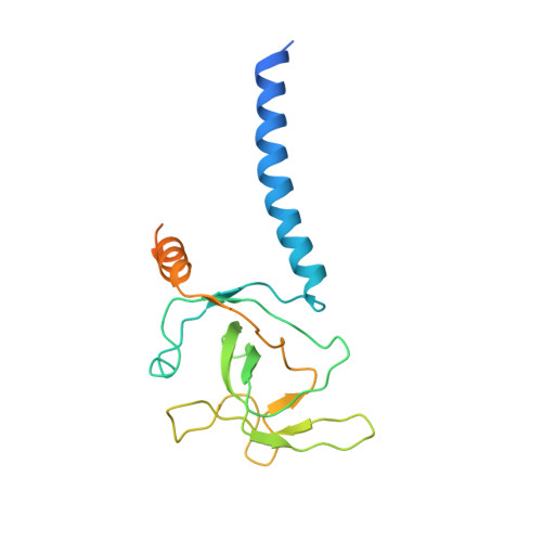
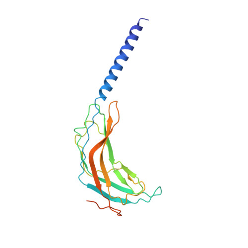
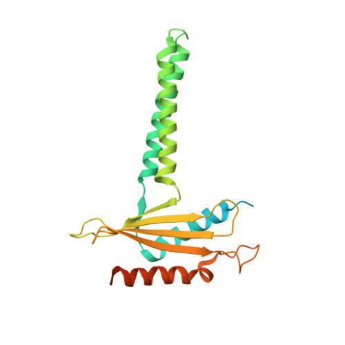
    The signal peptidase complex (SPC) is an essential membrane complex in the endoplasmic reticulum (ER), where it removes signal peptides (SPs) from a large variety of secretory pre-proteins with exquisite specificity. Although the determinants of this process have been established empirically, the molecular details of SP recognition and removal remain elusive. Here, we show that the human SPC exists in two functional paralogs with distinct proteolytic subunits. We determined the atomic structures of both paralogs using electron cryo-microscopy and structural proteomics. The active site is formed by a catalytic triad and abuts the ER membrane, where a transmembrane window collectively formed by all subunits locally thins the bilayer. Molecular dynamics simulations indicate that this unique architecture generates specificity for SPs based on the length of their hydrophobic segments.


  • Organizational Affiliation

    Structural Biochemistry, Bijvoet Centre for Biomolecular Research, Utrecht University, Universiteitsweg 99, 3584 CG, Utrecht, the Netherlands.


Macromolecules
Find similar proteins by:  (by identity cutoff)  |  3D Structure
Entity ID: 1
MoleculeChains Sequence LengthOrganismDetailsImage
Signal peptidase complex catalytic subunit SEC11A198Homo sapiensMutation(s): 0 
Gene Names: SEC11ASEC11L1SPC18SPCS4A
EC: 3.4.21.89
Membrane Entity: Yes 
UniProt & NIH Common Fund Data Resources
Find proteins for P67812 (Homo sapiens)
Explore P67812 
Go to UniProtKB:  P67812
PHAROS:  P67812
GTEx:  ENSG00000140612 
Entity Groups  
Sequence Clusters30% Identity50% Identity70% Identity90% Identity95% Identity100% Identity
UniProt GroupP67812
Sequence Annotations
Expand
  • Reference Sequence
Find similar proteins by:  (by identity cutoff)  |  3D Structure
Entity ID: 2
MoleculeChains Sequence LengthOrganismDetailsImage
Signal peptidase complex subunit 3201Homo sapiensMutation(s): 0 
Gene Names: SPCS3SPC22UNQ1841/PRO3567
EC: 3.4
Membrane Entity: Yes 
UniProt & NIH Common Fund Data Resources
Find proteins for P61009 (Homo sapiens)
Explore P61009 
Go to UniProtKB:  P61009
PHAROS:  P61009
GTEx:  ENSG00000129128 
Entity Groups  
Sequence Clusters30% Identity50% Identity70% Identity90% Identity95% Identity100% Identity
UniProt GroupP61009
Sequence Annotations
Expand
  • Reference Sequence
Find similar proteins by:  (by identity cutoff)  |  3D Structure
Entity ID: 3
MoleculeChains Sequence LengthOrganismDetailsImage
Signal peptidase complex subunit 2248Homo sapiensMutation(s): 0 
Gene Names: SPCS2KIAA0102SPC25
EC: 3.4
Membrane Entity: Yes 
UniProt & NIH Common Fund Data Resources
Find proteins for Q15005 (Homo sapiens)
Explore Q15005 
Go to UniProtKB:  Q15005
GTEx:  ENSG00000118363 
Entity Groups  
Sequence Clusters30% Identity50% Identity70% Identity90% Identity95% Identity100% Identity
UniProt GroupQ15005
Sequence Annotations
Expand
  • Reference Sequence
Find similar proteins by:  (by identity cutoff)  |  3D Structure
Entity ID: 4
MoleculeChains Sequence LengthOrganismDetailsImage
Signal peptidase complex subunit 1191Homo sapiensMutation(s): 0 
Gene Names: SPCS1SPC12HSPC033
EC: 3.4
Membrane Entity: Yes 
UniProt & NIH Common Fund Data Resources
Find proteins for Q9Y6A9 (Homo sapiens)
Explore Q9Y6A9 
Go to UniProtKB:  Q9Y6A9
PHAROS:  Q9Y6A9
GTEx:  ENSG00000114902 
Entity Groups  
Sequence Clusters30% Identity50% Identity70% Identity90% Identity95% Identity100% Identity
UniProt GroupQ9Y6A9
Sequence Annotations
Expand
  • Reference Sequence
Oligosaccharides

Help

Entity ID: 5
MoleculeChains Length2D Diagram Glycosylation3D Interactions
alpha-D-mannopyranose-(1-3)-[alpha-D-mannopyranose-(1-6)]beta-D-mannopyranose-(1-4)-2-acetamido-2-deoxy-beta-D-glucopyranose-(1-4)-2-acetamido-2-deoxy-beta-D-glucopyranose
E
5N-Glycosylation
Glycosylation Resources
GlyTouCan:  G22768VO
GlyCosmos:  G22768VO
GlyGen:  G22768VO
Experimental Data & Validation

Experimental Data

  • Method: ELECTRON MICROSCOPY
  • Resolution: 4.90 Å
  • Aggregation State: PARTICLE 
  • Reconstruction Method: SINGLE PARTICLE 
EM Software:
TaskSoftware PackageVersion
RECONSTRUCTIONRELION3.1
MODEL REFINEMENTPHENIX1.18.2

Structure Validation

View Full Validation Report


Entry History & Funding Information

Deposition Data


Funding OrganizationLocationGrant Number
European Research Council (ERC)Netherlands724425

Revision History  (Full details and data files)

  • Version 1.0: 2021-10-06
    Type: Initial release
  • Version 1.1: 2021-10-13
    Changes: Data collection
  • Version 1.2: 2021-10-20
    Changes: Data collection, Database references
  • Version 1.3: 2022-12-21
    Changes: Database references