7ONY | pdb_00007ony

Crystal structure of PBP3 from P. aeruginosa


Experimental Data Snapshot

  • Method: X-RAY DIFFRACTION
  • Resolution: 1.77 Å
  • R-Value Free: 
    0.213 (Depositor), 0.222 (DCC) 
  • R-Value Work: 
    0.180 (Depositor), 0.186 (DCC) 
  • R-Value Observed: 
    0.181 (Depositor) 

Starting Model: experimental
View more details

wwPDB Validation 3D Report Full Report

Validation slider image for 7ONY

This is version 1.2 of the entry. See complete history

Literature

Interaction Mode of the Novel Monobactam AIC499 Targeting Penicillin Binding Protein 3 of Gram-Negative Bacteria.

Freischem, S.Grimm, I.Lopez-Perez, A.Willbold, D.Klenke, B.Vuong, C.Dingley, A.J.Weiergraber, O.H.

(2021) Biomolecules 11

  • DOI: https://doi.org/10.3390/biom11071057
  • Primary Citation Related Structures: 
    7ONK, 7ONN, 7ONO, 7ONW, 7ONX, 7ONY, 7ONZ

  • PubMed Abstract: 

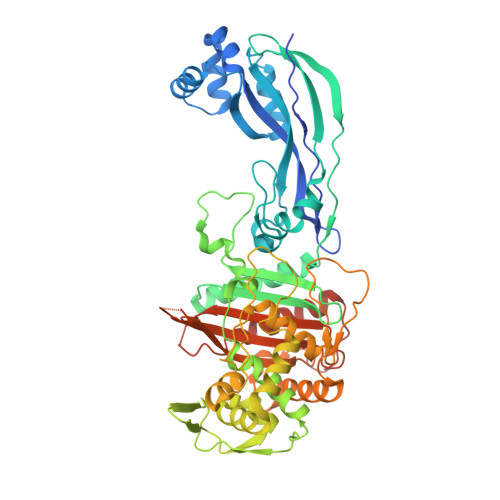
    Novel antimicrobial strategies are urgently required because of the rising threat of multi drug resistant bacterial strains and the infections caused by them. Among the available target structures, the so-called penicillin binding proteins are of particular interest, owing to their good accessibility in the periplasmic space, and the lack of homologous proteins in humans, reducing the risk of side effects of potential drugs. In this report, we focus on the interaction of the innovative β-lactam antibiotic AIC499 with penicillin binding protein 3 (PBP3) from Escherichia coli and Pseudomonas aeruginosa . This recently developed monobactam displays broad antimicrobial activity, against Gram-negative strains, and improved resistance to most classes of β-lactamases. By analyzing crystal structures of the respective complexes, we were able to explore the binding mode of AIC499 to its target proteins. In addition, the apo structures determined for PBP3, from P. aeruginosa and the catalytic transpeptidase domain of the E. coli orthologue, provide new insights into the dynamics of these proteins and the impact of drug binding.


  • Organizational Affiliation
    • Institute of Biological Information Processing (IBI-7: Structural Biochemistry) and Jülich Centre for Structural Biology (JuStruct), Forschungszentrum Jülich, 52425 Jülich, Germany.

Macromolecule Content 

  • Total Structure Weight: 59.11 kDa 
  • Atom Count: 4,233 
  • Modeled Residue Count: 505 
  • Deposited Residue Count: 531 
  • Unique protein chains: 1

Macromolecules

Find similar proteins by:|  3D Structure
Entity ID: 1
MoleculeChains  Sequence LengthOrganismDetailsImage
Peptidoglycan D,D-transpeptidase FtsI531Pseudomonas aeruginosa PAO1Mutation(s): 0 
Gene Names: ftsIpbpBPA4418
EC: 3.4.16.4
UniProt
Find proteins for G3XD46 (Pseudomonas aeruginosa (strain ATCC 15692 / DSM 22644 / CIP 104116 / JCM 14847 / LMG 12228 / 1C / PRS 101 / PAO1))
Explore G3XD46 
Go to UniProtKB:  G3XD46
Entity Groups
Sequence Clusters30% Identity50% Identity70% Identity90% Identity95% Identity100% Identity
UniProt GroupG3XD46
Sequence Annotations
Expand
Reference Sequence

Small Molecules

Ligands 3 Unique
IDChains Name / Formula / InChI Key2D Diagram3D Interactions
MES

Query on MES


Download:Ideal Coordinates CCD File
K [auth A],
L [auth A],
M [auth A]
2-(N-MORPHOLINO)-ETHANESULFONIC ACID
C6 H13 N O4 S
SXGZJKUKBWWHRA-UHFFFAOYSA-N
GOL

Query on GOL


Download:Ideal Coordinates CCD File
B [auth A]
C [auth A]
D [auth A]
E [auth A]
F [auth A]
B [auth A],
C [auth A],
D [auth A],
E [auth A],
F [auth A],
G [auth A],
H [auth A],
I [auth A],
J [auth A]
GLYCEROL
C3 H8 O3
PEDCQBHIVMGVHV-UHFFFAOYSA-N
DMS

Query on DMS


Download:Ideal Coordinates CCD File
N [auth A]DIMETHYL SULFOXIDE
C2 H6 O S
IAZDPXIOMUYVGZ-UHFFFAOYSA-N

Experimental Data & Validation

Experimental Data

  • Method: X-RAY DIFFRACTION
  • Resolution: 1.77 Å
  • R-Value Free:  0.213 (Depositor), 0.222 (DCC) 
  • R-Value Work:  0.180 (Depositor), 0.186 (DCC) 
  • R-Value Observed: 0.181 (Depositor) 
Space Group: C 1 2 1
Unit Cell:
Length ( Å )Angle ( ˚ )
a = 104.057α = 90
b = 124.995β = 122.494
c = 74.235γ = 90
Software Package:
Software NamePurpose
XDSdata reduction
Aimlessdata scaling
MOLREPphasing
PHENIXrefinement
PDB_EXTRACTdata extraction

Structure Validation

View Full Validation Report


Entry History 

Deposition Data

Revision History  (Full details and data files)

  • Version 1.0: 2021-08-04
    Type: Initial release
  • Version 1.1: 2021-09-22
    Changes: Data collection, Database references
  • Version 1.2: 2024-01-31
    Changes: Data collection, Refinement description