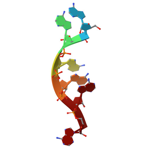
Cryo-EM structure of the cetacean morbillivirus nucleoprotein-RNA complex.
Zinzula, L., Beck, F., Klumpe, S., Bohn, S., Pfeifer, G., Bollschweiler, D., Nagy, I., Plitzko, J.M., Baumeister, W.(2021) J Struct Biol 213: 107750-107750
- PubMed: 34089875 Search on PubMed
- DOI: https://doi.org/10.1016/j.jsb.2021.107750
- Primary Citation Related Structures:
7OI3 - PubMed Abstract:
Cetacean morbillivirus (CeMV) is an emerging and highly infectious paramyxovirus that causes outbreaks in cetaceans and occasionally in pinnipeds, representing a major threat to biodiversity and conservation of endangered marine mammal populations in both hemispheres. As for all non-segmented, negative-sense, single-stranded RNA (ssRNA) viruses, the morbilliviral genome is enwrapped by thousands of nucleoprotein (N) protomers. Each bound to six ribonucleotides, N protomers assemble to form a helical ribonucleoprotein (RNP) complex that serves as scaffold for nucleocapsid formation and as template for viral replication and transcription. While the molecular details on RNP complexes elucidated in human measles virus (MeV) served as paradigm model for these processes in all members of the Morbillivirus genus, no structural information has been obtained from other morbilliviruses, nor has any CeMV structure been solved so far. We report the structure of the CeMV RNP complex, reconstituted in vitro upon binding of recombinant CeMV N to poly-adenine ssRNA hexamers and solved to 4.0 Å resolution by cryo-electron microscopy. In spite of the amino acid sequence similarity and consequently similar folding of the N protomer, the CeMV RNP complex exhibits different helical parameters as compared to previously reported MeV orthologs. The CeMV structure reveals exclusive interactions leading to more extensive protomer-RNA and protomer-protomer interfaces. We identified twelve residues, among those varying between CeMV strains, as putatively important for the stabilization of the RNP complex, which highlights the need to study the potential of CeMV N mutations that modulate nucleocapsid assembly to also affect viral phenotype and host adaptation.
- Max-Planck Institute of Biochemistry, Department of Molecular Structural Biology, Am Klopferspitz 18, 82152 Martinsried, Germany. Electronic address: zinzula@biochem.mpg.de.
Organizational Affiliation: