7OHD

CRYSTAL STRUCTURE OF FERRIC MURINE NEUROGLOBIN CDLESS MUTANT


Experimental Data Snapshot

  • Method: X-RAY DIFFRACTION
  • Resolution: 1.80 Å
  • R-Value Free: 0.211 
  • R-Value Work: 0.182 
  • R-Value Observed: 0.184 

wwPDB Validation   3D Report Full Report


Ligand Structure Quality Assessment 


This is version 1.2 of the entry. See complete history


Literature

Probing the Role of Murine Neuroglobin CDloop-D-Helix Unit in CO Ligand Binding and Structural Dynamics.

Exertier, C.Sebastiani, F.Freda, I.Gugole, E.Cerutti, G.Parisi, G.Montemiglio, L.C.Becucci, M.Viappiani, C.Bruno, S.Savino, C.Zamparelli, C.Anselmi, M.Abbruzzetti, S.Smulevich, G.Vallone, B.

(2022) ACS Chem Biol 17: 2099-2108

  • DOI: https://doi.org/10.1021/acschembio.2c00172
  • Primary Citation of Related Structures:  
    7OHD

  • PubMed Abstract: 

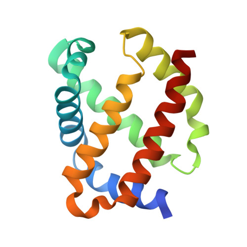
    We produced a neuroglobin variant, namely, Ngb CDless, with the excised CDloop- and D-helix, directly joining the C- and E-helices. The CDless variant retained bis-His hexacoordination, and we investigated the role of the CDloop-D-helix unit in controlling the CO binding and structural dynamics by an integrative approach based on X-ray crystallography, rapid mixing, laser flash photolysis, resonance Raman spectroscopy, and molecular dynamics simulations. Rapid mixing and laser flash photolysis showed that ligand affinity was unchanged with respect to the wild-type protein, albeit with increased on and off constants for rate-limiting heme iron hexacoordination by the distal His64. Accordingly, resonance Raman spectroscopy highlighted a more open distal pocket in the CO complex that, in agreement with MD simulations, likely involves His64 swinging inward and outward of the distal heme pocket. Ngb CDless displays a more rigid overall structure with respect to the wild type, abolishing the structural dynamics of the CDloop-D-helix hypothesized to mediate its signaling role, and it retains ligand binding control by distal His64. In conclusion, this mutant may represent a tool to investigate the involvement of CDloop-D-helix in neuroprotective signaling in a cellular or animal model.


  • Organizational Affiliation

    Dipartimento di Scienze Biochimiche "A. Rossi Fanelli", Sapienza, Università di Roma, Piazzale A. Moro 5, I-00185 Rome, Italy.


Macromolecules
Find similar proteins by:  (by identity cutoff)  |  3D Structure
Entity ID: 1
MoleculeChains Sequence LengthOrganismDetailsImage
Neuroglobin
A, B
139Mus musculusMutation(s): 1 
Gene Names: Ngb
UniProt
Find proteins for Q9ER97 (Mus musculus)
Explore Q9ER97 
Go to UniProtKB:  Q9ER97
Entity Groups  
Sequence Clusters30% Identity50% Identity70% Identity90% Identity95% Identity100% Identity
UniProt GroupQ9ER97
Sequence Annotations
Expand
  • Reference Sequence
Small Molecules
Ligands 5 Unique
IDChains Name / Formula / InChI Key2D Diagram3D Interactions
HEM
Query on HEM

Download Ideal Coordinates CCD File 
C [auth A],
J [auth B]
PROTOPORPHYRIN IX CONTAINING FE
C34 H32 Fe N4 O4
KABFMIBPWCXCRK-RGGAHWMASA-L
BTB
Query on BTB

Download Ideal Coordinates CCD File 
I [auth A]2-[BIS-(2-HYDROXY-ETHYL)-AMINO]-2-HYDROXYMETHYL-PROPANE-1,3-DIOL
C8 H19 N O5
OWMVSZAMULFTJU-UHFFFAOYSA-N
SO4
Query on SO4

Download Ideal Coordinates CCD File 
D [auth A],
F [auth A]
SULFATE ION
O4 S
QAOWNCQODCNURD-UHFFFAOYSA-L
GOL
Query on GOL

Download Ideal Coordinates CCD File 
E [auth A],
G [auth A],
L [auth B],
M [auth B]
GLYCEROL
C3 H8 O3
PEDCQBHIVMGVHV-UHFFFAOYSA-N
IPA
Query on IPA

Download Ideal Coordinates CCD File 
H [auth A],
K [auth B]
ISOPROPYL ALCOHOL
C3 H8 O
KFZMGEQAYNKOFK-UHFFFAOYSA-N
Experimental Data & Validation

Experimental Data

  • Method: X-RAY DIFFRACTION
  • Resolution: 1.80 Å
  • R-Value Free: 0.211 
  • R-Value Work: 0.182 
  • R-Value Observed: 0.184 
  • Space Group: P 63
Unit Cell:
Length ( Å )Angle ( ˚ )
a = 95.941α = 90
b = 95.941β = 90
c = 58.56γ = 120
Software Package:
Software NamePurpose
REFMACrefinement
PDB_EXTRACTdata extraction
xia2data reduction
MOLREPphasing
Cootmodel building
XSCALEdata scaling

Structure Validation

View Full Validation Report


Ligand Structure Quality Assessment 


Entry History & Funding Information

Deposition Data


Funding OrganizationLocationGrant Number
H2020 Marie Curie Actions of the European CommissionEuropean Union637295

Revision History  (Full details and data files)

  • Version 1.0: 2022-07-13
    Type: Initial release
  • Version 1.1: 2022-09-28
    Changes: Database references
  • Version 1.2: 2024-01-31
    Changes: Data collection, Refinement description