7OAT

Structural basis for targeted p97 remodelling by ASPL as prerequisite for p97 trimethylation by METTL21D


Experimental Data Snapshot

  • Method: X-RAY DIFFRACTION
  • Resolution: 3.00 Å
  • R-Value Free: 0.270 
  • R-Value Work: 0.225 
  • R-Value Observed: 0.228 

wwPDB Validation   3D Report Full Report


Ligand Structure Quality Assessment 


This is version 1.2 of the entry. See complete history


Literature

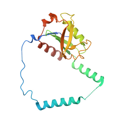
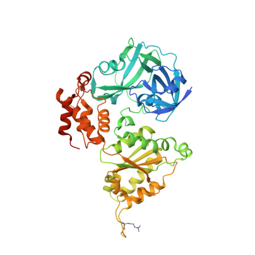
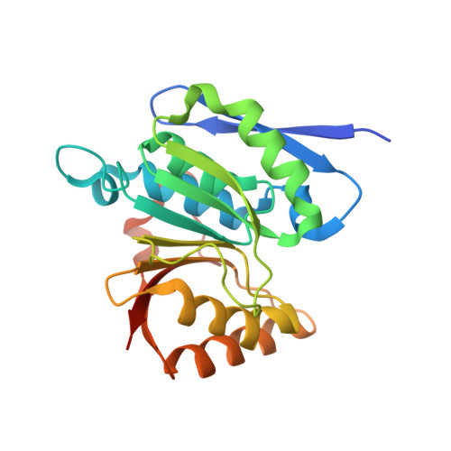
Structural remodeling of AAA+ ATPase p97 by adaptor protein ASPL facilitates posttranslational methylation by METTL21D.

Petrovic, S.Roske, Y.Rami, B.Phan, M.H.Q.Panakova, D.Heinemann, U.

(2023) Proc Natl Acad Sci U S A 120: e2208941120-e2208941120

  • DOI: https://doi.org/10.1073/pnas.2208941120
  • Primary Citation of Related Structures:  
    7OAS, 7OAT

  • PubMed Abstract: 

    p97 is an essential AAA+ ATPase that extracts and unfolds substrate proteins from membranes and protein complexes. Through its mode of action, p97 contributes to various cellular processes, such as membrane fusion, ER-associated protein degradation, DNA repair, and many others. Diverse p97 functions and protein interactions are regulated by a large number of adaptor proteins. Alveolar soft part sarcoma locus (ASPL) is a unique adaptor protein that regulates p97 by disassembling functional p97 hexamers to smaller entities. An alternative mechanism to regulate the activity and interactions of p97 is by posttranslational modifications (PTMs). Although more than 140 PTMs have been identified in p97, only a handful of those have been described in detail. Here we present structural and biochemical data to explain how the p97-remodeling adaptor protein ASPL enables the metastasis promoting methyltransferase METTL21D to bind and trimethylate p97 at a single lysine side chain, which is deeply buried inside functional p97 hexamers. The crystal structure of a heterotrimeric p97:ASPL:METTL21D complex in the presence of cofactors ATP and S-adenosyl homocysteine reveals how structural remodeling by ASPL exposes the crucial lysine residue of p97 to facilitate its trimethylation by METTL21D. The structure also uncovers a role of the second region of homology (SRH) present in the first ATPase domain of p97 in binding of a modifying enzyme to the AAA+ ATPase. Investigation of this interaction in the human, fish, and plant reveals fine details on the mechanism and significance of p97 trimethylation by METTL21D across different organisms.


  • Organizational Affiliation

    Max Delbrück Center for Molecular Medicine 13125, Berlin, Germany.


Macromolecules
Find similar proteins by:  (by identity cutoff)  |  3D Structure
Entity ID: 1
MoleculeChains Sequence LengthOrganismDetailsImage
Tether containing UBX domain for GLUT4190Homo sapiensMutation(s): 0 
Gene Names: ASPSCR1ASPLRCC17TUGUBXD9UBXN9
UniProt & NIH Common Fund Data Resources
Find proteins for Q9BZE9 (Homo sapiens)
Explore Q9BZE9 
Go to UniProtKB:  Q9BZE9
PHAROS:  Q9BZE9
GTEx:  ENSG00000169696 
Entity Groups  
Sequence Clusters30% Identity50% Identity70% Identity90% Identity95% Identity100% Identity
UniProt GroupQ9BZE9
Sequence Annotations
Expand
  • Reference Sequence
Find similar proteins by:  (by identity cutoff)  |  3D Structure
Entity ID: 2
MoleculeChains Sequence LengthOrganismDetailsImage
Transitional endoplasmic reticulum ATPase481Homo sapiensMutation(s): 0 
Gene Names: VCP
EC: 3.6.4.6
UniProt & NIH Common Fund Data Resources
Find proteins for P55072 (Homo sapiens)
Explore P55072 
Go to UniProtKB:  P55072
PHAROS:  P55072
GTEx:  ENSG00000165280 
Entity Groups  
Sequence Clusters30% Identity50% Identity70% Identity90% Identity95% Identity100% Identity
UniProt GroupP55072
Sequence Annotations
Expand
  • Reference Sequence
Find similar proteins by:  (by identity cutoff)  |  3D Structure
Entity ID: 3
MoleculeChains Sequence LengthOrganismDetailsImage
Protein-lysine methyltransferase METTL21D224Homo sapiensMutation(s): 0 
Gene Names: VCPKMTC14orf138METTL21D
EC: 2.1.1
UniProt & NIH Common Fund Data Resources
Find proteins for Q9H867 (Homo sapiens)
Explore Q9H867 
Go to UniProtKB:  Q9H867
PHAROS:  Q9H867
GTEx:  ENSG00000100483 
Entity Groups  
Sequence Clusters30% Identity50% Identity70% Identity90% Identity95% Identity100% Identity
UniProt GroupQ9H867
Sequence Annotations
Expand
  • Reference Sequence
Small Molecules
Ligands 4 Unique
IDChains Name / Formula / InChI Key2D Diagram3D Interactions
ATP
Query on ATP

Download Ideal Coordinates CCD File 
F [auth B]ADENOSINE-5'-TRIPHOSPHATE
C10 H16 N5 O13 P3
ZKHQWZAMYRWXGA-KQYNXXCUSA-N
SAH (Subject of Investigation/LOI)
Query on SAH

Download Ideal Coordinates CCD File 
I [auth C]S-ADENOSYL-L-HOMOCYSTEINE
C14 H20 N6 O5 S
ZJUKTBDSGOFHSH-WFMPWKQPSA-N
EDO
Query on EDO

Download Ideal Coordinates CCD File 
D [auth B],
E [auth B],
H [auth C],
J [auth C]
1,2-ETHANEDIOL
C2 H6 O2
LYCAIKOWRPUZTN-UHFFFAOYSA-N
MG
Query on MG

Download Ideal Coordinates CCD File 
G [auth B]MAGNESIUM ION
Mg
JLVVSXFLKOJNIY-UHFFFAOYSA-N
Modified Residues  1 Unique
IDChains TypeFormula2D DiagramParent
M3L
Query on M3L
B
L-PEPTIDE LINKINGC9 H21 N2 O2LYS
Experimental Data & Validation

Experimental Data

  • Method: X-RAY DIFFRACTION
  • Resolution: 3.00 Å
  • R-Value Free: 0.270 
  • R-Value Work: 0.225 
  • R-Value Observed: 0.228 
  • Space Group: P 1 21 1
Unit Cell:
Length ( Å )Angle ( ˚ )
a = 54.258α = 90
b = 69.622β = 94.41
c = 140.175γ = 90
Software Package:
Software NamePurpose
BUSTERrefinement
XDSdata reduction
XSCALEdata scaling
PDB_EXTRACTdata extraction
PHASERphasing

Structure Validation

View Full Validation Report


Ligand Structure Quality Assessment 


Entry History & Funding Information

Deposition Data


Funding OrganizationLocationGrant Number
German Research Foundation (DFG)GermanySFB740 TPB1

Revision History  (Full details and data files)

  • Version 1.0: 2022-08-03
    Type: Initial release
  • Version 1.1: 2023-02-15
    Changes: Database references
  • Version 1.2: 2024-02-07
    Changes: Data collection, Refinement description