


Crystal structure of Human Menin in complex with Fragment 21
Groves, M.R., Gao, K.To be published.
Experimental Data Snapshot
Starting Model: experimental
View more details
Entity ID: 1 | |||||
|---|---|---|---|---|---|
| Molecule | Chains | Sequence Length | Organism | Details | Image |
| Isoform 2 of Menin | 505 | Homo sapiens | Mutation(s): 0 Gene Names: MEN1, SCG2 | 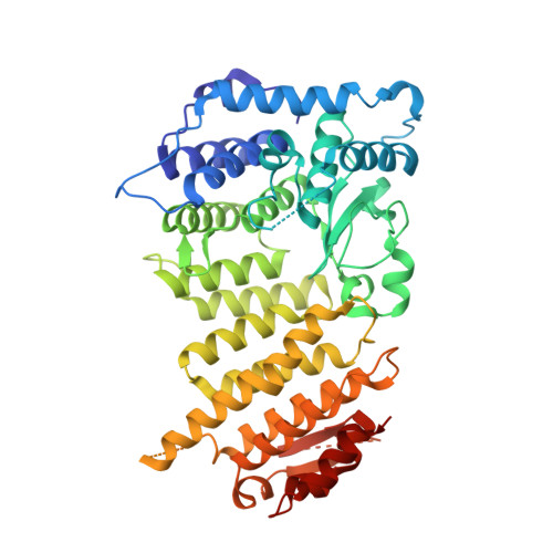 | |
UniProt & NIH Common Fund Data Resources | |||||
PHAROS: O00255 GTEx: ENSG00000133895 | |||||
Entity Groups | |||||
| Sequence Clusters | 30% Identity50% Identity70% Identity90% Identity95% Identity100% Identity | ||||
| UniProt Group | O00255 | ||||
Sequence AnnotationsExpand | |||||
Reference Sequence | |||||
| Ligands 3 Unique | |||||
|---|---|---|---|---|---|
| ID | Chains | Name / Formula / InChI Key | 2D Diagram | 3D Interactions | |
| V6K (Subject of Investigation/LOI) Download:Ideal Coordinates CCD File | B [auth A] | (~{E})-2-cyano-3-(4-hydroxyphenyl)-~{N}-(2-morpholin-4-ylethyl)prop-2-enamide C16 H19 N3 O3 FJCWZDJYNREWJS-SDNWHVSQSA-N |  | ||
| GOL Download:Ideal Coordinates CCD File | D [auth A] | GLYCEROL C3 H8 O3 PEDCQBHIVMGVHV-UHFFFAOYSA-N |  | ||
| EDO Download:Ideal Coordinates CCD File | C [auth A] E [auth A] F [auth A] G [auth A] H [auth A] | 1,2-ETHANEDIOL C2 H6 O2 LYCAIKOWRPUZTN-UHFFFAOYSA-N |  | ||
| Length ( Å ) | Angle ( ˚ ) |
|---|---|
| a = 107.399 | α = 90 |
| b = 107.399 | β = 90 |
| c = 107.903 | γ = 90 |
| Software Name | Purpose |
|---|---|
| XDS | data reduction |
| Aimless | data scaling |
| PHENIX | refinement |
| PDB_EXTRACT | data extraction |
| PHENIX | phasing |


