Insight on molecular pathogenesis and pharmacochaperoning potential in phosphomannomutase 2 deficiency, provided by novel human phosphomannomutase 2 structures.
Briso-Montiano, A., Del Cano-Ochoa, F., Vilas, A., Velazquez-Campoy, A., Rubio, V., Perez, B., Ramon-Maiques, S.(2022) J Inherit Metab Dis 45: 318-333
- PubMed: 34859900 Search on PubMed
- DOI: https://doi.org/10.1002/jimd.12461
- Primary Citation Related Structures:
7O0C, 7O1B, 7O4G, 7O58, 7O5Z - PubMed Abstract:
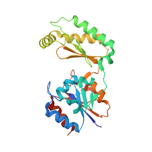
Phosphomannomutase 2 (PMM2) deficiency, the most frequent congenital disorder of glycosylation (PMM2-CDG), is a severe condition, which has no cure. Due to the identification of destabilizing mutations, our group aims at increasing residual activity in PMM2-CDG patients, searching for pharmacochaperones. Detailed structural knowledge of hPMM2 might help identify variants amenable to pharmacochaperoning. hPMM2 structural information is limited to one incomplete structure deposited in the Protein Databank without associated publication, which lacked ligands and residues from a crucial loop. Here we report five complete crystal structures of hPMM2, three for wild-type and two for the p.Thr237Met variant frequently found among Spanish PMM2-CDG patients, free and bound to the essential activator glucose-1,6-bisphosphate (Glc-1,6-P 2 ). In the hPMM2 homodimer, each subunit has a different conformation, reflecting movement of the distal core domain relative to the dimerization cap domain, supporting an opening/closing process during catalysis. Two Mg 2+ ions bind to the core domain, one catalytic and one structural. In the cap domain, the site for Glc-1,6-P 2 is well delineated, while a Cl - ion binding at the intersubunit interface is predicted to strengthen dimerization. Patient-found amino acid substitutions are nonhomogeneously distributed throughout hPMM2, reflecting differential functional or structural importance for various parts of the protein. We classify 93 of 101 patient-reported single amino acid variants according to five potential pathogenetic mechanism affecting folding of the core and cap domains, linker 2 flexibility, dimerization, activator binding, and catalysis. We propose that ~80% and ~50% of the respective core and cap domains substitutions are potential candidates for pharmacochaperoning treatment.
- Centro de Biología Molecular Severo Ochoa (CBMSO), Universidad Autónoma de Madrid and Consejo Superior de Investigaciones Científicas, Cantoblanco, Madrid, Spain.
Organizational Affiliation: