7NPU

MycP5-free ESX-5 inner membrane complex, state I


Experimental Data Snapshot

  • Method: ELECTRON MICROSCOPY
  • Resolution: 4.48 Å
  • Aggregation State: PARTICLE 
  • Reconstruction Method: SINGLE PARTICLE 

wwPDB Validation   3D Report Full Report


This is version 1.0 of the entry. See complete history


Literature

Structure and dynamics of a mycobacterial type VII secretion system.

Bunduc, C.M.Fahrenkamp, D.Wald, J.Ummels, R.Bitter, W.Houben, E.N.G.Marlovits, T.C.

(2021) Nature 593: 445-448

  • DOI: https://doi.org/10.1038/s41586-021-03517-z
  • Primary Citation of Related Structures:  
    7NP7, 7NPR, 7NPS, 7NPT, 7NPU, 7NPV

  • PubMed Abstract: 

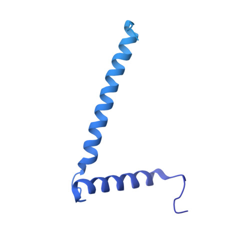
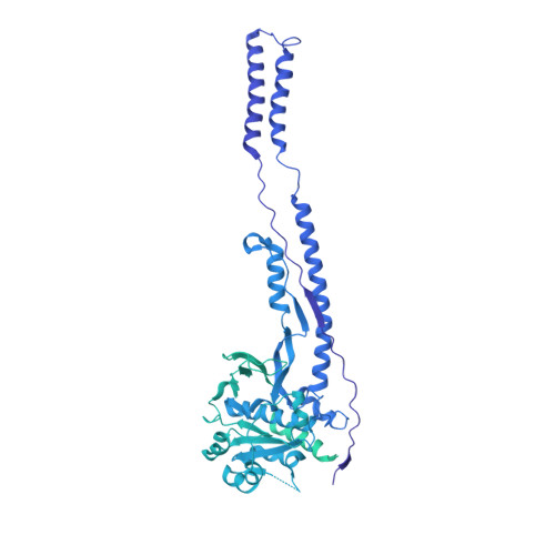
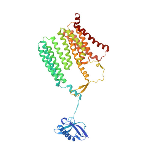
    Mycobacterium tuberculosis is the cause of one of the most important infectious diseases in humans, which leads to 1.4 million deaths every year 1 . Specialized protein transport systems-known as type VII secretion systems (T7SSs)-are central to the virulence of this pathogen, and are also crucial for nutrient and metabolite transport across the mycobacterial cell envelope 2,3 . Here we present the structure of an intact T7SS inner-membrane complex of M. tuberculosis. We show how the 2.32-MDa ESX-5 assembly, which contains 165 transmembrane helices, is restructured and stabilized as a trimer of dimers by the MycP 5 protease. A trimer of MycP 5 caps a central periplasmic dome-like chamber that is formed by three EccB 5 dimers, with the proteolytic sites of MycP 5 facing towards the cavity. This chamber suggests a central secretion and processing conduit. Complexes without MycP 5 show disruption of the EccB 5 periplasmic assembly and increased flexibility, which highlights the importance of MycP 5 for complex integrity. Beneath the EccB 5 -MycP 5 chamber, dimers of the EccC 5 ATPase assemble into three bundles of four transmembrane helices each, which together seal the potential central secretion channel. Individual cytoplasmic EccC 5 domains adopt two distinctive conformations that probably reflect different secretion states. Our work suggests a previously undescribed mechanism of protein transport and provides a structural scaffold to aid in the development of drugs against this major human pathogen.


  • Organizational Affiliation

    Centre for Structural Systems Biology, Hamburg, Germany.


Macromolecules
Find similar proteins by:  (by identity cutoff)  |  3D Structure
Entity ID: 1
MoleculeChains Sequence LengthOrganismDetailsImage
ESX-5 secretion system ATPase EccB5506Mycobacterium tuberculosis H37RvMutation(s): 0 
Gene Names: eccB5Rv1782
EC: 3.6
Membrane Entity: Yes 
UniProt
Find proteins for P9WNQ9 (Mycobacterium tuberculosis (strain ATCC 25618 / H37Rv))
Explore P9WNQ9 
Go to UniProtKB:  P9WNQ9
Entity Groups  
Sequence Clusters30% Identity50% Identity70% Identity90% Identity95% Identity100% Identity
UniProt GroupP9WNQ9
Sequence Annotations
Expand
  • Reference Sequence
Find similar proteins by:  (by identity cutoff)  |  3D Structure
Entity ID: 2
MoleculeChains Sequence LengthOrganismDetailsImage
ESX-5 secretion system protein EccC51,391Mycobacterium tuberculosis H37RvMutation(s): 0 
Gene Names: eccC5Rv1783
Membrane Entity: Yes 
UniProt
Find proteins for P9WNA5 (Mycobacterium tuberculosis (strain ATCC 25618 / H37Rv))
Explore P9WNA5 
Go to UniProtKB:  P9WNA5
Entity Groups  
Sequence Clusters30% Identity50% Identity70% Identity90% Identity95% Identity100% Identity
UniProt GroupP9WNA5
Sequence Annotations
Expand
  • Reference Sequence
Find similar proteins by:  (by identity cutoff)  |  3D Structure
Entity ID: 3
MoleculeChains Sequence LengthOrganismDetailsImage
ESX-5 secretion system protein EccD5503Mycobacterium tuberculosis H37RvMutation(s): 0 
Gene Names: eccD5Rv1795
Membrane Entity: Yes 
UniProt
Find proteins for P9WNP9 (Mycobacterium tuberculosis (strain ATCC 25618 / H37Rv))
Explore P9WNP9 
Go to UniProtKB:  P9WNP9
Entity Groups  
Sequence Clusters30% Identity50% Identity70% Identity90% Identity95% Identity100% Identity
UniProt GroupP9WNP9
Sequence Annotations
Expand
  • Reference Sequence
Experimental Data & Validation

Experimental Data

  • Method: ELECTRON MICROSCOPY
  • Resolution: 4.48 Å
  • Aggregation State: PARTICLE 
  • Reconstruction Method: SINGLE PARTICLE 
EM Software:
TaskSoftware PackageVersion
MODEL REFINEMENTISOLDE1.0.1
MODEL REFINEMENTPHENIX1.18

Structure Validation

View Full Validation Report


Entry History & Funding Information

Deposition Data


Funding OrganizationLocationGrant Number
German Research Foundation (DFG)GermanyINST 152/774-1 FUGG
Netherlands Organisation for Scientific Research (NWO)Netherlands864.12.006
H2020 Marie Curie Actions of the European CommissionEuropean Union101030373
German Research Foundation (DFG)GermanyFA1518/2-1

Revision History  (Full details and data files)

  • Version 1.0: 2021-06-02
    Type: Initial release