7NI3 | pdb_00007ni3

CRYSTAL STRUCTURE OF NATIVE HUMAN MYELOPEROXIDASE IN COMPLEX WITH CPD 3


Experimental Data Snapshot

  • Method: X-RAY DIFFRACTION
  • Resolution: 2.10 Å
  • R-Value Free: 
    0.224 (Depositor), 0.235 (DCC) 
  • R-Value Work: 
    0.175 (Depositor), 0.182 (DCC) 
  • R-Value Observed: 
    0.177 (Depositor) 

Starting Model: experimental
View more details

wwPDB Validation 3D Report Full Report

Validation slider image for 7NI3

Ligand Structure Quality Assessment 


This is version 1.6 of the entry. See complete history

Literature

Discovery of AZD4831, a Mechanism-Based Irreversible Inhibitor of Myeloperoxidase, As a Potential Treatment for Heart Failure with Preserved Ejection Fraction.

Inghardt, T.Antonsson, T.Ericsson, C.Hovdal, D.Johannesson, P.Johansson, C.Jurva, U.Kajanus, J.Kull, B.Michaelsson, E.Pettersen, A.Sjogren, T.Sorensen, H.Westerlund, K.Lindstedt, E.L.

(2022) J Med Chem 65: 11485-11496

  • DOI: https://doi.org/10.1021/acs.jmedchem.1c02141
  • Primary Citation Related Structures: 
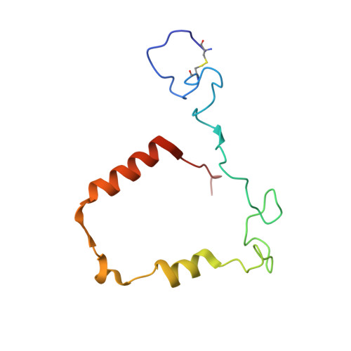
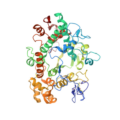
    7NI1, 7NI3

  • PubMed Abstract: 

    Myeloperoxidase is a promising therapeutic target for treatment of patients suffering from heart failure with preserved ejection fraction (HFpEF). We aimed to discover a covalent myeloperoxidase inhibitor with high selectivity for myeloperoxidase over thyroid peroxidase, limited penetration of the blood-brain barrier, and pharmacokinetics suitable for once-daily oral administration at low dose. Structure-activity relationship, biophysical, and structural studies led to prioritization of four compounds for in-depth safety and pharmacokinetic studies in animal models. One compound (AZD4831) progressed to clinical studies on grounds of high potency (IC 50 , 1.5 nM in vitro ) and selectivity (>450-fold vs thyroid peroxidase in vitro ), the mechanism of irreversible inhibition, and the safety profile. Following phase 1 studies in healthy volunteers and a phase 2a study in patients with HFpEF, a phase 2 b /3 efficacy study of AZD4831 in patients with HFpEF started in 2021.


  • Organizational Affiliation
    • Research and Early Development, Cardiovascular, Renal and Metabolism, BioPharmaceuticals R&D, AstraZeneca, 431 50 Mölndal, Gothenburg, Sweden.

Macromolecule Content 

  • Total Structure Weight: 135.12 kDa 
  • Atom Count: 10,213 
  • Modeled Residue Count: 1,136 
  • Deposited Residue Count: 1,142 
  • Unique protein chains: 2

Macromolecules

Find similar proteins by:|  3D Structure
Entity ID: 1
MoleculeChains  Sequence LengthOrganismDetailsImage
Myeloperoxidase
A, B
105Homo sapiensMutation(s): 0 
EC: 1.11.2.2
UniProt & NIH Common Fund Data Resources
Find proteins for P05164 (Homo sapiens)
Explore P05164 
Go to UniProtKB:  P05164
PHAROS:  P05164
GTEx:  ENSG00000005381 
Entity Groups
Sequence Clusters30% Identity50% Identity70% Identity90% Identity95% Identity100% Identity
UniProt GroupP05164
Sequence Annotations
Expand
Reference Sequence
Find similar proteins by:|  3D Structure
Entity ID: 2
MoleculeChains  Sequence LengthOrganismDetailsImage
Myeloperoxidase
C, D
466Homo sapiensMutation(s): 0 
EC: 1.11.2.2
UniProt & NIH Common Fund Data Resources
Find proteins for P05164 (Homo sapiens)
Explore P05164 
Go to UniProtKB:  P05164
PHAROS:  P05164
GTEx:  ENSG00000005381 
Entity Groups
Sequence Clusters30% Identity50% Identity70% Identity90% Identity95% Identity100% Identity
UniProt GroupP05164
Glycosylation
Glycosylation Sites: 3Go to GlyGen: P05164-1
Sequence Annotations
Expand
Reference Sequence

Oligosaccharides

Help  
Entity ID: 3
MoleculeChains Length2D Diagram GlycosylationD Interactions
beta-D-mannopyranose-(1-4)-2-acetamido-2-deoxy-beta-D-glucopyranose-(1-4)-[alpha-L-fucopyranose-(1-6)]2-acetamido-2-deoxy-beta-D-glucopyranose
E, F
4N-Glycosylation
Glycosylation Resources
GlyTouCan: G32152BH
GlyCosmos: G32152BH
GlyGen: G32152BH

Small Molecules

Ligands 6 Unique
IDChains Name / Formula / InChI Key2D Diagram3D Interactions
HEM

Query on HEM


Download:Ideal Coordinates CCD File
G [auth A],
J [auth B]
PROTOPORPHYRIN IX CONTAINING FE
C34 H32 Fe N4 O4
KABFMIBPWCXCRK-RGGAHWMASA-L
UE8
(Subject of Investigation/LOI)

Query on UE8


Download:Ideal Coordinates CCD File
M [auth C],
S [auth D]
2-sulfanylidene-3-[(2R)-tetrahydro-2-furanylmethyl]-1,2,3,7-tetrahydro-6H-purin-6-one
C10 H12 N4 O2 S
RSPDBEVKURKEII-ZCFIWIBFSA-N
NAG

Query on NAG


Download:Ideal Coordinates CCD File
P [auth C],
Q [auth C],
U [auth D],
V [auth D]
2-acetamido-2-deoxy-beta-D-glucopyranose
C8 H15 N O6
OVRNDRQMDRJTHS-FMDGEEDCSA-N
BMA

Query on BMA


Download:Ideal Coordinates CCD File
R [auth C],
T [auth D]
beta-D-mannopyranose
C6 H12 O6
WQZGKKKJIJFFOK-RWOPYEJCSA-N
CA

Query on CA


Download:Ideal Coordinates CCD File
N [auth C],
W [auth D]
CALCIUM ION
Ca
BHPQYMZQTOCNFJ-UHFFFAOYSA-N
CL

Query on CL


Download:Ideal Coordinates CCD File
H [auth A],
I [auth B],
K [auth B],
L [auth C],
O [auth C]
CHLORIDE ION
Cl
VEXZGXHMUGYJMC-UHFFFAOYSA-M
Modified Residues  1 Unique
IDChains TypeFormula2D DiagramParent
CSO
Query on CSO
C, D
L-PEPTIDE LINKINGC3 H7 N O3 SCYS

Experimental Data & Validation

Experimental Data

  • Method: X-RAY DIFFRACTION
  • Resolution: 2.10 Å
  • R-Value Free:  0.224 (Depositor), 0.235 (DCC) 
  • R-Value Work:  0.175 (Depositor), 0.182 (DCC) 
  • R-Value Observed: 0.177 (Depositor) 
Space Group: P 1 21 1
Unit Cell:
Length ( Å )Angle ( ˚ )
a = 92.66α = 90
b = 63.64β = 97.15
c = 111.03γ = 90
Software Package:
Software NamePurpose
XDSdata reduction
STARANISOdata scaling
PHASERphasing
BUSTERrefinement

Structure Validation

View Full Validation Report


Ligand Structure Quality Assessment 


Entry History 

Deposition Data

Revision History  (Full details and data files)

  • Version 1.0: 2022-02-23
    Type: Initial release
  • Version 1.1: 2022-04-20
    Changes: Database references
  • Version 1.2: 2022-08-31
    Changes: Database references
  • Version 1.3: 2022-09-07
    Changes: Database references
  • Version 1.4: 2022-09-21
    Changes: Database references
  • Version 1.5: 2024-01-31
    Changes: Data collection, Refinement description
  • Version 1.6: 2024-11-20
    Changes: Structure summary