7NH9

structure of the full-length CmaX protein


Experimental Data Snapshot

  • Method: ELECTRON MICROSCOPY
  • Resolution: 3.03 Å
  • Aggregation State: PARTICLE 
  • Reconstruction Method: SINGLE PARTICLE 

wwPDB Validation   3D Report Full Report


This is version 1.2 of the entry. See complete history


Literature

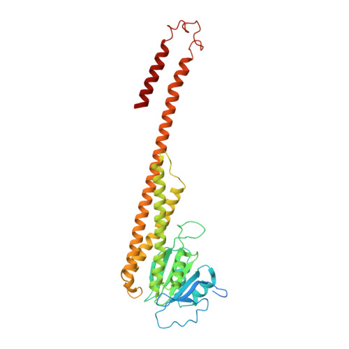
Structural and biochemical characterization of a novel ZntB (CmaX) transporter protein from Pseudomonas aeruginosa.

Stetsenko, A.Stehantsev, P.Dranenko, N.O.Gelfand, M.S.Guskov, A.

(2021) Int J Biol Macromol 184: 760-767

  • DOI: https://doi.org/10.1016/j.ijbiomac.2021.06.130
  • Primary Citation of Related Structures:  
    7NH9

  • PubMed Abstract: 

    The 2-TM-GxN family of membrane proteins is widespread in prokaryotes and plays an important role in transport of divalent cations. The canonical signature motif, which is also a selectivity filter, has a composition of Gly-Met-Asn. Some members though deviate from this composition, however no data are available as to whether this has any functional implications. Here we report the functional and structural analysis of CmaX protein from a pathogenic Pseudomonas aeruginosa bacterium, which has a Gly-Ile-Asn signature motif. CmaX readily transports Zn 2+ , Mg 2+ , Cd 2+ , Ni 2+ and Co 2+ ions, but it does not utilize proton-symport as does ZntB from Escherichia coli. Together with the bioinformatics analysis, our data suggest that deviations from the canonical signature motif do not reveal any changes in substrate selectivity or transport and easily alter in course of evolution.


  • Organizational Affiliation

    Groningen Biomolecular Sciences and Biotechnology Institute, University of Groningen, the Netherlands.


Macromolecules
Find similar proteins by:  (by identity cutoff)  |  3D Structure
Entity ID: 1
MoleculeChains Sequence LengthOrganismDetailsImage
CmaX protein
A, B, C, D, E
366Pseudomonas aeruginosa PAO1Mutation(s): 0 
Gene Names: cmaXPA1773
Membrane Entity: Yes 
UniProt
Find proteins for G3XD00 (Pseudomonas aeruginosa (strain ATCC 15692 / DSM 22644 / CIP 104116 / JCM 14847 / LMG 12228 / 1C / PRS 101 / PAO1))
Explore G3XD00 
Go to UniProtKB:  G3XD00
Entity Groups  
Sequence Clusters30% Identity50% Identity70% Identity90% Identity95% Identity100% Identity
UniProt GroupG3XD00
Sequence Annotations
Expand
  • Reference Sequence
Experimental Data & Validation

Experimental Data

  • Method: ELECTRON MICROSCOPY
  • Resolution: 3.03 Å
  • Aggregation State: PARTICLE 
  • Reconstruction Method: SINGLE PARTICLE 

Structure Validation

View Full Validation Report


Entry History & Funding Information

Deposition Data


Funding OrganizationLocationGrant Number
Netherlands Organisation for Scientific Research (NWO)740.018.011

Revision History  (Full details and data files)

  • Version 1.0: 2021-07-07
    Type: Initial release
  • Version 1.1: 2021-07-14
    Changes: Database references
  • Version 1.2: 2024-05-01
    Changes: Data collection, Database references