7NEQ

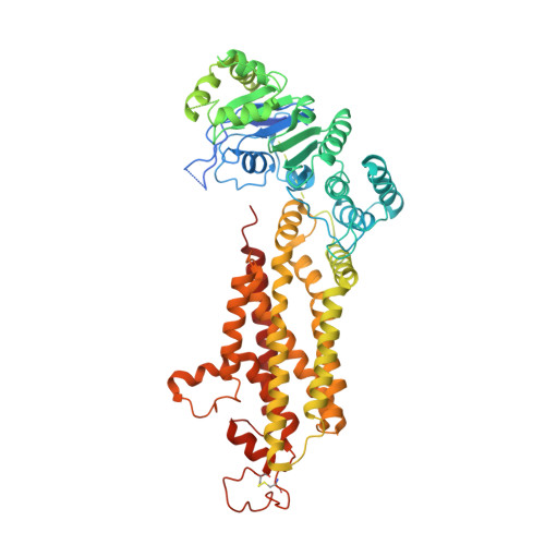
Structure of tariquidar-bound ABCG2


Experimental Data Snapshot

  • Method: ELECTRON MICROSCOPY
  • Resolution: 3.12 Å
  • Aggregation State: PARTICLE 
  • Reconstruction Method: SINGLE PARTICLE 

wwPDB Validation   3D Report Full Report


This is version 1.1 of the entry. See complete history


Literature

Structural Basis of Drug Recognition by the Multidrug Transporter ABCG2.

Kowal, J.Ni, D.Jackson, S.M.Manolaridis, I.Stahlberg, H.Locher, K.P.

(2021) J Mol Biol 433: 166980-166980

  • DOI: https://doi.org/10.1016/j.jmb.2021.166980
  • Primary Citation of Related Structures:  
    7NEQ, 7NEZ, 7NFD

  • PubMed Abstract: 

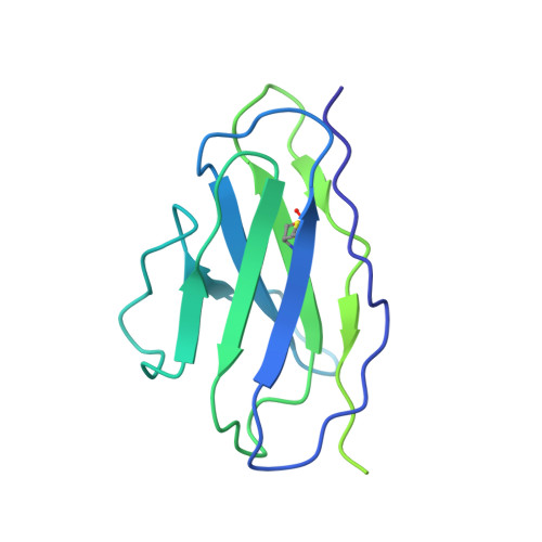
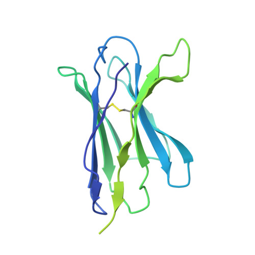
    ABCG2 is an ATP-binding cassette (ABC) transporter whose function affects the pharmacokinetics of drugs and contributes to multidrug resistance of cancer cells. While its interaction with the endogenous substrate estrone-3-sulfate (E 1 S) has been elucidated at a structural level, the recognition and recruitment of exogenous compounds is not understood at sufficiently high resolution. Here we present three cryo-EM structures of nanodisc-reconstituted, human ABCG2 bound to anticancer drugs tariquidar, topotecan and mitoxantrone. To enable structural insight at high resolution, we used Fab fragments of the ABCG2-specific monoclonal antibody 5D3, which binds to the external side of the transporter but does not interfere with drug-induced stimulation of ATPase activity. We observed that the binding pocket of ABCG2 can accommodate a single tariquidar molecule in a C-shaped conformation, similar to one of the two tariquidar molecules bound to ABCB1, where tariquidar acts as an inhibitor. We also found single copies of topotecan and mitoxantrone bound between key phenylalanine residues. Mutagenesis experiments confirmed the functional importance of two residues in the binding pocket, F439 and N436. Using 3D variability analyses, we found a correlation between substrate binding and reduced dynamics of the nucleotide binding domains (NBDs), suggesting a structural explanation for drug-induced ATPase stimulation. Our findings provide additional insight into how ABCG2 differentiates between inhibitors and substrates and may guide a rational design of new modulators and substrates.


  • Organizational Affiliation

    Institute of Molecular Biology and Biophysics, ETH Zurich, Switzerland. Electronic address: julia.kowal@mol.biol.ethz.ch.


Macromolecules
Find similar proteins by:  (by identity cutoff)  |  3D Structure
Entity ID: 1
MoleculeChains Sequence LengthOrganismDetailsImage
ATP-binding cassette sub-family G member 2A,
F [auth B]
655Homo sapiensMutation(s): 0 
Gene Names: ABCG2ABCPBCRPBCRP1MXR
EC: 7.6.2.2
Membrane Entity: Yes 
UniProt & NIH Common Fund Data Resources
Find proteins for Q9UNQ0 (Homo sapiens)
Explore Q9UNQ0 
Go to UniProtKB:  Q9UNQ0
PHAROS:  Q9UNQ0
GTEx:  ENSG00000118777 
Entity Groups  
Sequence Clusters30% Identity50% Identity70% Identity90% Identity95% Identity100% Identity
UniProt GroupQ9UNQ0
Glycosylation
Glycosylation Sites: 1Go to GlyGen: Q9UNQ0-1
Sequence Annotations
Expand
  • Reference Sequence
Find similar proteins by:  (by identity cutoff)  |  3D Structure
Entity ID: 2
MoleculeChains Sequence LengthOrganismDetailsImage
5D3(Fab) light chain variable domainB [auth C],
D [auth E]
214Mus musculusMutation(s): 0 
Entity Groups  
Sequence Clusters30% Identity50% Identity70% Identity90% Identity95% Identity100% Identity
Sequence Annotations
Expand
  • Reference Sequence
Find similar proteins by:  (by identity cutoff)  |  3D Structure
Entity ID: 3
MoleculeChains Sequence LengthOrganismDetailsImage
5D3(Fab) heavy chain variable domainC [auth D],
E [auth F]
221Mus musculusMutation(s): 0 
Entity Groups  
Sequence Clusters30% Identity50% Identity70% Identity90% Identity95% Identity100% Identity
Sequence Annotations
Expand
  • Reference Sequence
Small Molecules
Ligands 4 Unique
IDChains Name / Formula / InChI Key2D Diagram3D Interactions
R1H (Subject of Investigation/LOI)
Query on R1H

Download Ideal Coordinates CCD File 
K [auth B]tariquidar
C38 H38 N4 O6
LGGHDPFKSSRQNS-UHFFFAOYSA-N
U9N
Query on U9N

Download Ideal Coordinates CCD File 
H [auth A][(2~{S})-3-[2-azanylethoxy(oxidanyl)phosphoryl]oxy-2-decanoyloxy-propyl] octadecanoate
C33 H66 N O8 P
IQGMGIHASGWWFZ-HKBQPEDESA-N
CLR
Query on CLR

Download Ideal Coordinates CCD File 
I [auth A],
J [auth A],
M [auth B]
CHOLESTEROL
C27 H46 O
HVYWMOMLDIMFJA-DPAQBDIFSA-N
NAG
Query on NAG

Download Ideal Coordinates CCD File 
G [auth A],
L [auth B]
2-acetamido-2-deoxy-beta-D-glucopyranose
C8 H15 N O6
OVRNDRQMDRJTHS-FMDGEEDCSA-N
Binding Affinity Annotations 
IDSourceBinding Affinity
R1H BindingDB:  7NEQ IC50: min: 183, max: 6223 (nM) from 10 assay(s)
EC50: min: 10, max: 100 (nM) from 2 assay(s)
Experimental Data & Validation

Experimental Data

  • Method: ELECTRON MICROSCOPY
  • Resolution: 3.12 Å
  • Aggregation State: PARTICLE 
  • Reconstruction Method: SINGLE PARTICLE 
EM Software:
TaskSoftware PackageVersion
RECONSTRUCTIONRELION3.1

Structure Validation

View Full Validation Report


Entry History & Funding Information

Deposition Data


Funding OrganizationLocationGrant Number
Swiss National Science FoundationSwitzerlandTransCure

Revision History  (Full details and data files)

  • Version 1.0: 2021-04-21
    Type: Initial release
  • Version 1.1: 2021-05-12
    Changes: Database references