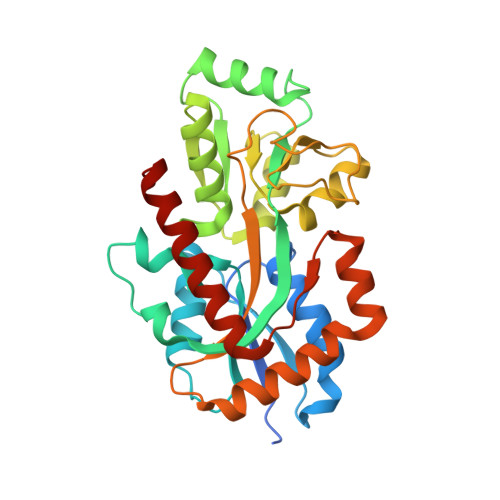
7NDS

Crystal structure of TphC in a closed conformation


Experimental Data Snapshot

  • Method: X-RAY DIFFRACTION
  • Resolution: 2.40 Å
  • R-Value Free: 0.217 
  • R-Value Work: 0.178 
  • R-Value Observed: 0.180 

Starting Model: experimental
View more details

wwPDB Validation   3D Report Full Report


This is version 1.2 of the entry. See complete history


Literature

Structural basis of terephthalate recognition by solute binding protein TphC.

Gautom, T.Dheeman, D.Levy, C.Butterfield, T.Alvarez Gonzalez, G.Le Roy, P.Caiger, L.Fisher, K.Johannissen, L.Dixon, N.

(2021) Nat Commun 12: 6244-6244

  • DOI: https://doi.org/10.1038/s41467-021-26508-0
  • Primary Citation of Related Structures:  
    7NDR, 7NDS

  • PubMed Abstract: 

    Biological degradation of Polyethylene terephthalate (PET) plastic and assimilation of the corresponding monomers ethylene glycol and terephthalate (TPA) into central metabolism offers an attractive route for bio-based molecular recycling and bioremediation applications. A key step is the cellular uptake of the non-permeable TPA into bacterial cells which has been shown to be dependent upon the presence of the key tphC gene. However, little is known from a biochemical and structural perspective about the encoded solute binding protein, TphC. Here, we report the biochemical and structural characterisation of TphC in both open and TPA-bound closed conformations. This analysis demonstrates the narrow ligand specificity of TphC towards aromatic para-substituted dicarboxylates, such as TPA and closely related analogues. Further phylogenetic and genomic context analysis of the tph genes reveals homologous operons as a genetic resource for future biotechnological and metabolic engineering efforts towards circular plastic bio-economy solutions.


  • Organizational Affiliation

    Manchester Institute of Biotechnology (MIB) and Department of Chemistry, The University of Manchester, Manchester, UK.


Macromolecules
Find similar proteins by:  (by identity cutoff)  |  3D Structure
Entity ID: 1
MoleculeChains Sequence LengthOrganismDetailsImage
Tripartite tricarboxylate transporter substrate binding protein314Comamonas sp.Mutation(s): 0 
Gene Names: E2578_22390
UniProt
Find proteins for A0A5N7XFM8 (Comamonas sp)
Explore A0A5N7XFM8 
Go to UniProtKB:  A0A5N7XFM8
Entity Groups  
Sequence Clusters30% Identity50% Identity70% Identity90% Identity95% Identity100% Identity
UniProt GroupA0A5N7XFM8
Sequence Annotations
Expand
  • Reference Sequence
Small Molecules
Ligands 1 Unique
IDChains Name / Formula / InChI Key2D Diagram3D Interactions
UB7
Query on UB7

Download Ideal Coordinates CCD File 
B [auth A]terephthalic acid
C8 H6 O4
KKEYFWRCBNTPAC-UHFFFAOYSA-N
Experimental Data & Validation

Experimental Data

  • Method: X-RAY DIFFRACTION
  • Resolution: 2.40 Å
  • R-Value Free: 0.217 
  • R-Value Work: 0.178 
  • R-Value Observed: 0.180 
  • Space Group: C 2 2 21
Unit Cell:
Length ( Å )Angle ( ˚ )
a = 69.25α = 90
b = 150.06β = 90
c = 58.57γ = 90
Software Package:
Software NamePurpose
PHENIXrefinement
xia2data reduction
xia2data scaling
PHASERphasing

Structure Validation

View Full Validation Report


Entry History & Funding Information

Deposition Data

  • Released Date: 2021-11-03 
  • Deposition Author(s): Levy, C.

Funding OrganizationLocationGrant Number
Commonwealth Scholarship Commission (United Kingdom)United KingdomINCN-2018-57

Revision History  (Full details and data files)

  • Version 1.0: 2021-11-03
    Type: Initial release
  • Version 1.1: 2021-11-17
    Changes: Data collection, Database references, Structure summary
  • Version 1.2: 2024-01-31
    Changes: Data collection, Refinement description