7NDF | pdb_00007ndf

Crystal structure of nanobody Nb_MsbA#1 in complex with the nucleotide binding domain of MsbA


Experimental Data Snapshot

  • Method: X-RAY DIFFRACTION
  • Resolution: 2.10 Å
  • R-Value Free: 
    0.244 (Depositor), 0.247 (DCC) 
  • R-Value Work: 
    0.205 (Depositor), 0.209 (DCC) 
  • R-Value Observed: 
    0.207 (Depositor) 

Starting Model: experimental
View more details

wwPDB Validation 3D Report Full Report

Validation slider image for 7NDF

This is version 1.3 of the entry. See complete history

Literature

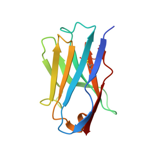
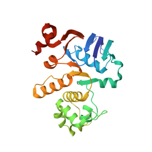
The ABC transporter MsbA adopts the wide inward-open conformation in E. coli cells.

Galazzo, L.Meier, G.Januliene, D.Parey, K.De Vecchis, D.Striednig, B.Hilbi, H.Schafer, L.V.Kuprov, I.Moeller, A.Bordignon, E.Seeger, M.A.

(2022) Sci Adv 8: eabn6845-eabn6845

  • DOI: https://doi.org/10.1126/sciadv.abn6845
  • Primary Citation Related Structures: 
    7NDF, 7PH2, 7PH3, 7PH4, 7PH7

  • PubMed Abstract: 

    Membrane proteins are currently investigated after detergent extraction from native cellular membranes and reconstitution into artificial liposomes or nanodiscs, thereby removing them from their physiological environment. However, to truly understand the biophysical properties of membrane proteins in a physiological environment, they must be investigated within living cells. Here, we used a spin-labeled nanobody to interrogate the conformational cycle of the ABC transporter MsbA by double electron-electron resonance. Unexpectedly, the wide inward-open conformation of MsbA, commonly considered a nonphysiological state, was found to be prominently populated in Escherichia coli cells. Molecular dynamics simulations revealed that extensive lateral portal opening is essential to provide access of its large natural substrate core lipid A to the binding cavity. Our work paves the way to investigate the conformational landscape of membrane proteins in cells.


  • Organizational Affiliation
    • Faculty of Chemistry and Biochemistry, Ruhr University Bochum, 44801 Bochum, Germany.

Macromolecule Content 

  • Total Structure Weight: 78.93 kDa 
  • Atom Count: 5,712 
  • Modeled Residue Count: 707 
  • Deposited Residue Count: 722 
  • Unique protein chains: 2

Macromolecules

Find similar proteins by:|  3D Structure
Entity ID: 1
MoleculeChains  Sequence LengthOrganismDetailsImage
Nb_MsbA 1A [auth C],
B [auth D]
116Vicugna pacosMutation(s): 0 
Find similar proteins by:|  3D Structure
Entity ID: 2
MoleculeChains  Sequence LengthOrganismDetailsImage
Lipid A ABC transporter ATP-binding protein/permease MsbAC [auth A],
D [auth B]
245Escherichia coliMutation(s): 0 
Gene Names: msbAHIR41_000141
EC: 7.5.2.6
UniProt
Find proteins for P60752 (Escherichia coli (strain K12))
Explore P60752 
Go to UniProtKB:  P60752
Entity Groups
Sequence Clusters30% Identity50% Identity70% Identity90% Identity95% Identity100% Identity
UniProt GroupP60752
Sequence Annotations
Expand
Reference Sequence

Experimental Data & Validation

Experimental Data

  • Method: X-RAY DIFFRACTION
  • Resolution: 2.10 Å
  • R-Value Free:  0.244 (Depositor), 0.247 (DCC) 
  • R-Value Work:  0.205 (Depositor), 0.209 (DCC) 
  • R-Value Observed: 0.207 (Depositor) 
Space Group: P 21 21 21
Unit Cell:
Length ( Å )Angle ( ˚ )
a = 63.53α = 90
b = 99.3β = 90
c = 142.45γ = 90
Software Package:
Software NamePurpose
PHENIXrefinement
XDSdata reduction
XSCALEdata scaling
PHASERphasing

Structure Validation

View Full Validation Report


Entry History 

& Funding Information

Deposition Data


Funding OrganizationLocationGrant Number
Swiss National Science FoundationSwitzerland310030_188817

Revision History  (Full details and data files)

  • Version 1.0: 2022-03-02
    Type: Initial release
  • Version 1.1: 2022-10-26
    Changes: Database references
  • Version 1.2: 2024-01-31
    Changes: Data collection, Refinement description
  • Version 1.3: 2024-10-16
    Changes: Structure summary