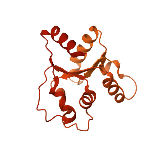
7NAK

Cryo-EM structure of activated human SARM1 in complex with NMN and 1AD (TIR:1AD)


Experimental Data Snapshot

  • Method: ELECTRON MICROSCOPY
  • Resolution: 2.90 Å
  • Aggregation State: PARTICLE 
  • Reconstruction Method: SINGLE PARTICLE 

wwPDB Validation   3D Report Full Report


This is version 1.2 of the entry. See complete history


Literature

Structural basis of SARM1 activation, substrate recognition, and inhibition by small molecules.

Shi, Y.Kerry, P.S.Nanson, J.D.Bosanac, T.Sasaki, Y.Krauss, R.Saikot, F.K.Adams, S.E.Mosaiab, T.Masic, V.Mao, X.Rose, F.Vasquez, E.Furrer, M.Cunnea, K.Brearley, A.Gu, W.Luo, Z.Brillault, L.Landsberg, M.J.DiAntonio, A.Kobe, B.Milbrandt, J.Hughes, R.O.Ve, T.

(2022) Mol Cell 82: 1643

  • DOI: https://doi.org/10.1016/j.molcel.2022.03.007
  • Primary Citation of Related Structures:  
    7NAG, 7NAH, 7NAI, 7NAJ, 7NAK, 7NAL

  • PubMed Abstract: 

    The NADase SARM1 (sterile alpha and TIR motif containing 1) is a key executioner of axon degeneration and a therapeutic target for several neurodegenerative conditions. We show that a potent SARM1 inhibitor undergoes base exchange with the nicotinamide moiety of nicotinamide adenine dinucleotide (NAD + ) to produce the bona fide inhibitor 1AD. We report structures of SARM1 in complex with 1AD, NAD + mimetics and the allosteric activator nicotinamide mononucleotide (NMN). NMN binding triggers reorientation of the armadillo repeat (ARM) domains, which disrupts ARM:TIR interactions and leads to formation of a two-stranded TIR domain assembly. The active site spans two molecules in these assemblies, explaining the requirement of TIR domain self-association for NADase activity and axon degeneration. Our results reveal the mechanisms of SARM1 activation and substrate binding, providing rational avenues for the design of new therapeutics targeting SARM1.


  • Organizational Affiliation

    Institute for Glycomics, Griffith University, Southport, QLD 4222, Australia.


Macromolecules
Find similar proteins by:  (by identity cutoff)  |  3D Structure
Entity ID: 1
MoleculeChains Sequence LengthOrganismDetailsImage
NAD(+) hydrolase SARM1697Homo sapiensMutation(s): 0 
Gene Names: SARM1KIAA0524SAMD2SARM
EC: 3.2.2.6 (PDB Primary Data), 3.2.2 (PDB Primary Data)
UniProt & NIH Common Fund Data Resources
Find proteins for Q6SZW1 (Homo sapiens)
Explore Q6SZW1 
Go to UniProtKB:  Q6SZW1
PHAROS:  Q6SZW1
GTEx:  ENSG00000004139 
Entity Groups  
Sequence Clusters30% Identity50% Identity70% Identity90% Identity95% Identity100% Identity
UniProt GroupQ6SZW1
Sequence Annotations
Expand
  • Reference Sequence
Experimental Data & Validation

Experimental Data

  • Method: ELECTRON MICROSCOPY
  • Resolution: 2.90 Å
  • Aggregation State: PARTICLE 
  • Reconstruction Method: SINGLE PARTICLE 
EM Software:
TaskSoftware PackageVersion
RECONSTRUCTIONcryoSPARC3.1

Structure Validation

View Full Validation Report


Entry History & Funding Information

Deposition Data


Funding OrganizationLocationGrant Number
National Health and Medical Research Council (NHMRC, Australia)Australia--
Australian Research Council (ARC)Australia--

Revision History  (Full details and data files)

  • Version 1.0: 2022-03-23
    Type: Initial release
  • Version 1.1: 2022-04-06
    Changes: Database references
  • Version 1.2: 2022-05-18
    Changes: Database references