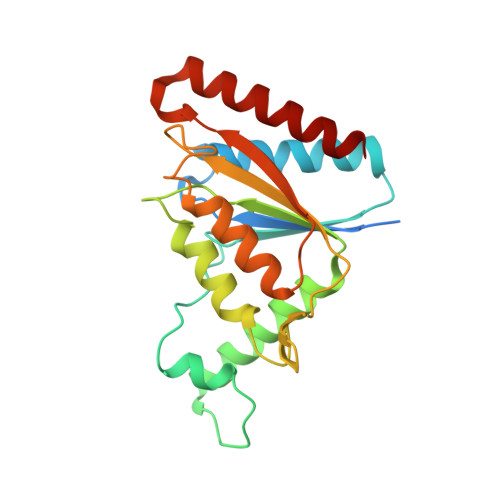
7N2X | pdb_00007n2x

The crystal structure of an FMN-dependent NADH:quinone oxidoreductase, AzoR from Escherichia coli


Experimental Data Snapshot

  • Method: X-RAY DIFFRACTION
  • Resolution: 1.70 Å
  • R-Value Free: 
    0.190 (Depositor), 0.194 (DCC) 
  • R-Value Work: 
    0.165 (Depositor), 0.169 (DCC) 
  • R-Value Observed: 
    0.167 (Depositor) 

Starting Model: experimental
View more details

wwPDB Validation 3D Report Full Report

Validation slider image for 7N2X

Ligand Structure Quality Assessment 


This is version 1.2 of the entry. See complete history. 

Literature

Ligand Binding Remodels the Oligomeric State and Active Site of Klebsiella pneumoniae Azoreductase.

Hitchings, R., Ghosh, A., Arcinas, A.J., Almo, S.C., Kelly, L., Ryan, A.

(2026) Biochemistry 65: 3236-3246

  • DOI: https://doi.org/10.1021/acs.biochem.6c00568
  • Primary Citation Related Structures: 
    6DXP, 7N2W, 7N2X

  • PubMed Abstract: 

    Azo dyes are widespread in the environment, and humans ingest a range of these dyes present in food and drugs. The human gut microbiome significantly impacts the fate of ingested azo dyes primarily through the reduction of the azo bond (R-N = N-R'). Flavin-dependent azoreductases have been considered the key enzymatic drivers of this process. To challenge this paradigm, we constructed a sequence similarity network of known azoreductases and focused on AzoA of Klebsiella pneumoniae, a central representative within the gut microbiome cluster. Unexpectedly, K. pneumoniae AzoA displayed relatively low in vitro activity against a range of azo compounds. While the physiological implications remain unclear, these findings are consistent with the possibility that nonenzymatic mechanisms contribute substantially to azo compound reduction in the gut. Furthermore, structural characterization of AzoA, as a holoenzyme and in its ligand-bound state, revealed a previously unrecognized ligand-coupled mechanism involving remodeling of the active site and oligomeric state.


  • Organizational Affiliation: 
    • Department of Systems and Computational Biology, Albert Einstein College of Medicine, Bronx, New York10461, United States.

Macromolecule Content 

  • Total Structure Weight: 25.12 kDa 
  • Atom Count: 1,781 
  • Modeled Residue Count: 201 
  • Deposited Residue Count: 223 
  • Unique protein chains: 1

Macromolecules

Find similar proteins by:|  3D Structure
Entity ID: 1
MoleculeChains  Sequence LengthOrganismDetailsImage
FMN-dependent NADH:quinone oxidoreductase223Escherichia coli O157:H7Mutation(s): 0 
Gene Names: azoR, Z2315, ECs2014
EC: 1.6.5 (PDB Primary Data), 1.7.1.17 (PDB Primary Data)
UniProt
Find proteins for P41407 (Escherichia coli (strain K12))
Explore P41407 
Go to UniProtKB:  P41407
Entity Groups
Sequence Clusters30% Identity50% Identity70% Identity90% Identity95% Identity100% Identity
UniProt GroupP41407
Sequence Annotations
Expand
Reference Sequence

Small Molecules

Ligands 4 Unique
IDChains Name / Formula / InChI Key2D Diagram3D Interactions
FMN

Query on FMN


Download:Ideal Coordinates CCD File
B [auth A]FLAVIN MONONUCLEOTIDE
C17 H21 N4 O9 P
FVTCRASFADXXNN-SCRDCRAPSA-N
P4G

Query on P4G


Download:Ideal Coordinates CCD File
E [auth A]1-ETHOXY-2-(2-ETHOXYETHOXY)ETHANE
C8 H18 O3
RRQYJINTUHWNHW-UHFFFAOYSA-N
DHA

Query on DHA


Download:Ideal Coordinates CCD File
D [auth A]2-AMINO-ACRYLIC ACID
C3 H5 N O2
UQBOJOOOTLPNST-UHFFFAOYSA-N
EDO

Query on EDO


Download:Ideal Coordinates CCD File
C [auth A],
F [auth A],
G [auth A]
1,2-ETHANEDIOL
C2 H6 O2
LYCAIKOWRPUZTN-UHFFFAOYSA-N

Experimental Data & Validation

Experimental Data

  • Method: X-RAY DIFFRACTION
  • Resolution: 1.70 Å
  • R-Value Free:  0.190 (Depositor), 0.194 (DCC) 
  • R-Value Work:  0.165 (Depositor), 0.169 (DCC) 
  • R-Value Observed: 0.167 (Depositor) 
Space Group: P 4 21 2
Unit Cell:
Length ( Å )Angle ( ˚ )
a = 94.355α = 90
b = 94.355β = 90
c = 53.081γ = 90
Software Package:
Software NamePurpose
PHENIXrefinement
MOSFLMdata reduction
PHASERphasing
PDB_EXTRACTdata extraction
SCALAdata scaling

Structure Validation

View Full Validation Report


Ligand Structure Quality Assessment 


Entry History 

Deposition Data

Revision History  (Full details and data files)

  • Version 1.0: 2022-08-31
    Type: Initial release
  • Version 1.1: 2023-10-18
    Changes: Data collection, Refinement description
  • Version 1.2: 2026-10-28
    Changes: Database references, Refinement description, Structure summary