To Be, or Not to Be, an Inhibitor: A Comparison of Azole Interactions with and Oxidation by a Cytochrome P450 Enzyme.
Podgorski, M.N., Coleman, T., Giang, P.D., Wang, C.R., Bruning, J.B., Bernhardt, P.V., De Voss, J.J., Bell, S.G.(2022) Inorg Chem 61: 236-245
- PubMed: 34910500 Search on PubMed
- DOI: https://doi.org/10.1021/acs.inorgchem.1c02786
- Primary Citation Related Structures:
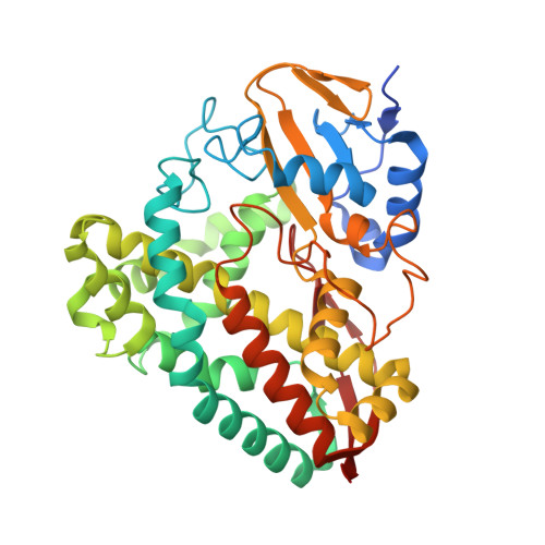
6U31, 7N14 - PubMed Abstract:
The cytochrome P450 (CYP) superfamily of heme monooxygenases is involved in a range of important chemical biotransformations across nature. Azole-containing molecules have been developed as drugs that bind to the heme center of these enzymes, inhibiting their function. The optical spectrum of CYP enzymes after the addition of these inhibitors is used to assess how the molecules bind. Here we use the bacterial CYP199A4 enzyme, from Rhodopseudomonas palustris HaA2, to compare how imidazolyl and triazolyl inhibitors bind to ferric and ferrous heme. 4-(Imidazol-1-yl)benzoic acid induced a red shift in the Soret wavelength (424 nm) in the ferric enzyme along with an increase and a decrease in the intensities of the δ and α bands, respectively. 4-(1 H -1,2,4-Triazol-1-yl)benzoic acid binds to CYP199A4 with a 10-fold lower affinity and induces a smaller red shift in the Soret band. The crystal structures of CYP199A4 with these two inhibitors confirmed that these differences in the optical spectra were due to coordination of the imidazolyl ligand to the ferric Fe, but the triazolyl inhibitor interacts with, rather than displaces, the ferric aqua ligand. Additional water molecules were present in the active site of 4-(1 H -1,2,4-triazol-1-yl)benzoic acid-bound CYP199A4. The space required to accommodate these additional water molecules in the active site necessitates changes in the position of the hydrophobic phenylalanine 298 residue. Upon reduction of the heme, the imidazole-based inhibitor Fe-N ligation was not retained. A 5-coordinate heme was also the predominant species in 4-(1 H -1,2,4-triazol-1-yl)benzoic acid-bound ferrous CYP199A4, but there was an obvious shoulder at 447 nm indicative of some degree of Fe-N coordination. Rather than inhibit CYP199A4, 4-(imidazol-1-yl)benzoic acid was a substrate and was oxidized to generate a metabolite derived from ring opening of the imidazolyl ring: 4-[[2-(formylamino)acetyl]amino]benzoic acid.
- Department of Chemistry, University of Adelaide, Adelaide, South Australia 5005, Australia.
Organizational Affiliation: