


Structural Basis for Branched Substrate Selectivity in a Ketoreductase from Ascaris suum
Dong, H., Chang, M.C.Y.(2021) ACS Catal 11: 8948-8955
Experimental Data Snapshot
Starting Model: experimental
View more details
(2021) ACS Catal 11: 8948-8955
Entity ID: 1 | |||||
|---|---|---|---|---|---|
| Molecule | Chains | Sequence Length | Organism | Details | Image |
| 3-hydroxyacyl-CoA dehydrogenase type-2 | 277 | Ascaris suum | Mutation(s): 0 EC: 1.1.1.53 (UniProt), 1.1.1.62 (UniProt) | 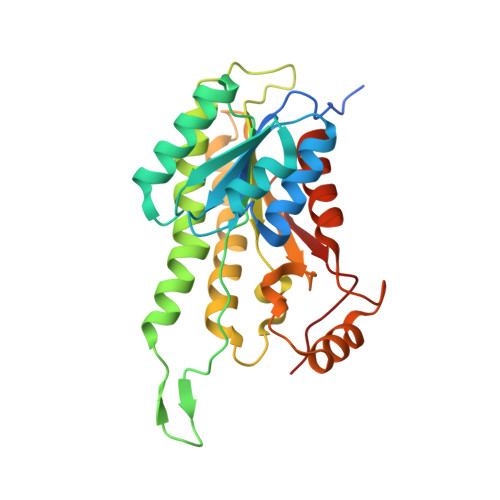 | |
UniProt | |||||
Entity Groups | |||||
| Sequence Clusters | 30% Identity50% Identity70% Identity90% Identity95% Identity100% Identity | ||||
| UniProt Group | F1L0H2 | ||||
Sequence AnnotationsExpand | |||||
Reference Sequence | |||||
| Ligands 1 Unique | |||||
|---|---|---|---|---|---|
| ID | Chains | Name / Formula / InChI Key | 2D Diagram | 3D Interactions | |
| NAD (Subject of Investigation/LOI) Download:Ideal Coordinates CCD File | C [auth A], D [auth B] | NICOTINAMIDE-ADENINE-DINUCLEOTIDE C21 H27 N7 O14 P2 BAWFJGJZGIEFAR-NNYOXOHSSA-N |  | ||
| Length ( Å ) | Angle ( ˚ ) |
|---|---|
| a = 128.542 | α = 90 |
| b = 54.942 | β = 131.039 |
| c = 89.27 | γ = 90 |
| Software Name | Purpose |
|---|---|
| PHENIX | refinement |
| XDS | data reduction |
| Aimless | data scaling |
| BALBES | phasing |
| Coot | model building |
| Funding Organization | Location | Grant Number |
|---|---|---|
| National Science Foundation (NSF, United States) | United States | CHE-1413862 |


