7MYX

Crystal structure of the PH domain (R86A) of Akt1


Experimental Data Snapshot

  • Method: X-RAY DIFFRACTION
  • Resolution: 1.39 Å
  • R-Value Free: 0.204 
  • R-Value Work: 0.175 
  • R-Value Observed: 0.178 

wwPDB Validation   3D Report Full Report


This is version 1.1 of the entry. See complete history


Literature

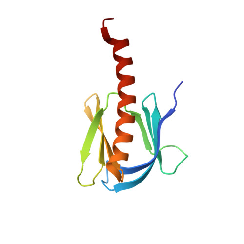
PH domain-mediated autoinhibition and oncogenic activation of Akt.

Bae, H.Viennet, T.Park, E.Chu, N.Salguero, A.Eck, M.J.Arthanari, H.Cole, P.A.

(2022) Elife 11

  • DOI: https://doi.org/10.7554/eLife.80148
  • Primary Citation of Related Structures:  
    7MYX

  • PubMed Abstract: 

    Akt is a Ser/Thr protein kinase that plays a central role in metabolism and cancer. Regulation of Akt's activity involves an autoinhibitory intramolecular interaction between its pleckstrin homology (PH) domain and its kinase domain that can be relieved by C-tail phosphorylation. PH domain mutant E17K Akt is a well-established oncogene. Previously, we reported that the conformation of autoinhibited Akt may be shifted by small molecule allosteric inhibitors limiting the mechanistic insights from existing X-ray structures that have relied on such compounds (Chu et al., 2020). Here, we discover unexpectedly that a single mutation R86A Akt exhibits intensified autoinhibitory features with enhanced PH domain-kinase domain affinity. Structural and biochemical analysis uncovers the importance of a key interaction network involving Arg86, Glu17, and Tyr18 that controls Akt conformation and activity. Our studies also shed light on the molecular basis for E17K Akt activation as an oncogenic driver.


  • Organizational Affiliation

    Department of Biological Chemistry and Molecular Pharmacology, Harvard Medical School, Boston, United States.


Macromolecules
Find similar proteins by:  (by identity cutoff)  |  3D Structure
Entity ID: 1
MoleculeChains Sequence LengthOrganismDetailsImage
RAC-alpha serine/threonine-protein kinase121Homo sapiensMutation(s): 1 
Gene Names: AKT1PKBRAC
EC: 2.7.11.1
UniProt & NIH Common Fund Data Resources
Find proteins for P31749 (Homo sapiens)
Explore P31749 
Go to UniProtKB:  P31749
PHAROS:  P31749
GTEx:  ENSG00000142208 
Entity Groups  
Sequence Clusters30% Identity50% Identity70% Identity90% Identity95% Identity100% Identity
UniProt GroupP31749
Sequence Annotations
Expand
  • Reference Sequence
Experimental Data & Validation

Experimental Data

  • Method: X-RAY DIFFRACTION
  • Resolution: 1.39 Å
  • R-Value Free: 0.204 
  • R-Value Work: 0.175 
  • R-Value Observed: 0.178 
  • Space Group: C 1 2 1
Unit Cell:
Length ( Å )Angle ( ˚ )
a = 84.786α = 90
b = 33.627β = 119.333
c = 42.352γ = 90
Software Package:
Software NamePurpose
PHENIXrefinement
XDSdata reduction
Cootmodel building
XDSdata scaling
PHASERphasing

Structure Validation

View Full Validation Report


Entry History 

Deposition Data

Revision History  (Full details and data files)

  • Version 1.0: 2022-11-09
    Type: Initial release
  • Version 1.1: 2023-10-18
    Changes: Data collection, Refinement description