7MYO

Cryo-EM structure of p110alpha in complex with p85alpha inhibited by BYL-719


Experimental Data Snapshot

  • Method: ELECTRON MICROSCOPY
  • Resolution: 2.92 Å
  • Aggregation State: PARTICLE 
  • Reconstruction Method: SINGLE PARTICLE 

wwPDB Validation   3D Report Full Report


This is version 1.1 of the entry. See complete history


Literature

Cryo-EM structures of PI3K alpha reveal conformational changes during inhibition and activation.

Liu, X.Yang, S.Hart, J.R.Xu, Y.Zou, X.Zhang, H.Zhou, Q.Xia, T.Zhang, Y.Yang, D.Wang, M.W.Vogt, P.K.

(2021) Proc Natl Acad Sci U S A 118

  • DOI: https://doi.org/10.1073/pnas.2109327118
  • Primary Citation of Related Structures:  
    7MYN, 7MYO

  • PubMed Abstract: 

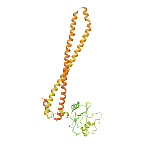
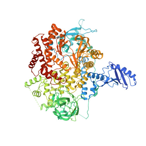
    Phosphoinositide 3-kinases (PI3Ks) are lipid kinases essential for growth and metabolism. Their aberrant activation is associated with many types of cancers. Here we used single-particle cryoelectron microscopy (cryo-EM) to determine three distinct conformations of full-length PI3Kα (p110α-p85α): the unliganded heterodimer PI3Kα, PI3Kα bound to the p110α-specific inhibitor BYL-719, and PI3Kα exposed to an activating phosphopeptide. The cryo-EM structures of unbound and of BYL-719-bound PI3Kα are in general accord with published crystal structures. Local deviations are presented and discussed. BYL-719 stabilizes the structure of PI3Kα, but three regions of low-resolution extra density remain and are provisionally assigned to the cSH2, BH, and SH3 domains of p85. One of the extra density regions is in contact with the kinase domain blocking access to the catalytic site. This conformational change indicates that the effects of BYL-719 on PI3Kα activity extend beyond competition with adenosine triphosphate (ATP). In unliganded PI3Kα, the DFG motif occurs in the "in" and "out" positions. In BYL-719-bound PI3Kα, only the DFG-in position, corresponding to the active conformation of the kinase, was observed. The phosphopeptide-bound structure of PI3Kα is composed of a stable core resolved at 3.8 Å. It contains all p110α domains except the adaptor-binding domain (ABD). The p85α domains, linked to the core through the ABD, are no longer resolved, implying that the phosphopeptide activates PI3Kα by fully releasing the niSH2 domain from binding to p110α. The structures presented here show the basal form of the full-length PI3Kα dimer and document conformational changes related to the activated and inhibited states.


  • Organizational Affiliation

    School of Pharmacy, Fudan University, Shanghai 201203, China.


Macromolecules
Find similar proteins by:  (by identity cutoff)  |  3D Structure
Entity ID: 1
MoleculeChains Sequence LengthOrganismDetailsImage
Phosphatidylinositol 3-kinase regulatory subunit alphaA [auth B]723Homo sapiensMutation(s): 0 
Gene Names: PIK3R1GRB1
UniProt & NIH Common Fund Data Resources
Find proteins for P27986 (Homo sapiens)
Explore P27986 
Go to UniProtKB:  P27986
PHAROS:  P27986
GTEx:  ENSG00000145675 
Entity Groups  
Sequence Clusters30% Identity50% Identity70% Identity90% Identity95% Identity100% Identity
UniProt GroupP27986
Sequence Annotations
Expand
  • Reference Sequence
Find similar proteins by:  (by identity cutoff)  |  3D Structure
Entity ID: 2
MoleculeChains Sequence LengthOrganismDetailsImage
Phosphatidylinositol 4,5-bisphosphate 3-kinase catalytic subunit alpha isoformB [auth A]1,096Homo sapiensMutation(s): 0 
Gene Names: PIK3CA
EC: 2.7.1.153 (PDB Primary Data), 2.7.11.1 (PDB Primary Data)
UniProt & NIH Common Fund Data Resources
Find proteins for P42336 (Homo sapiens)
Explore P42336 
Go to UniProtKB:  P42336
PHAROS:  P42336
GTEx:  ENSG00000121879 
Entity Groups  
Sequence Clusters30% Identity50% Identity70% Identity90% Identity95% Identity100% Identity
UniProt GroupP42336
Sequence Annotations
Expand
  • Reference Sequence
Small Molecules
Ligands 1 Unique
IDChains Name / Formula / InChI Key2D Diagram3D Interactions
1LT (Subject of Investigation/LOI)
Query on 1LT

Download Ideal Coordinates CCD File 
C [auth A](2S)-N~1~-{4-methyl-5-[2-(1,1,1-trifluoro-2-methylpropan-2-yl)pyridin-4-yl]-1,3-thiazol-2-yl}pyrrolidine-1,2-dicarboxamide
C19 H22 F3 N5 O2 S
STUWGJZDJHPWGZ-LBPRGKRZSA-N
Experimental Data & Validation

Experimental Data

  • Method: ELECTRON MICROSCOPY
  • Resolution: 2.92 Å
  • Aggregation State: PARTICLE 
  • Reconstruction Method: SINGLE PARTICLE 
EM Software:
TaskSoftware PackageVersion
MODEL REFINEMENTPHENIX1.19.2-4158
MODEL REFINEMENTCoot0.9.2-pre EL

Structure Validation

View Full Validation Report


Entry History & Funding Information

Deposition Data


Funding OrganizationLocationGrant Number
National Institutes of Health/National Cancer Institute (NIH/NCI)United StatesR35 CA197582
National Institutes of Health/National Cancer Institute (NIH/NCI)United StatesR50 CA243899
National Natural Science Foundation of China (NSFC)China81872915
National Natural Science Foundation of China (NSFC)China82073904
National Natural Science Foundation of China (NSFC)China81922071
National Natural Science Foundation of China (NSFC)China81773792
National Natural Science Foundation of China (NSFC)China81973373
National Natural Science Foundation of China (NSFC)China21704064
Ministry of Science and Technology (MoST, China)China2018ZX09735 001
Ministry of Science and Technology (MoST, China)China2018ZX09711002 002 005
Ministry of Science and Technology (MoST, China)China2018YFA0507000
Ministry of Science and Technology (MoST, China)China2019YFA0508800
Chinese Academy of SciencesChinaNNCAS 2017 1 CC

Revision History  (Full details and data files)

  • Version 1.0: 2021-11-10
    Type: Initial release
  • Version 1.1: 2021-11-24
    Changes: Database references