7MYB

Structure of proline utilization A with tetrahydrothiophene-2-carboxylate bound in the proline dehydrogenase active site


Experimental Data Snapshot

  • Method: X-RAY DIFFRACTION
  • Resolution: 1.52 Å
  • R-Value Free: 0.186 
  • R-Value Work: 0.164 
  • R-Value Observed: 0.165 

wwPDB Validation   3D Report Full Report


Ligand Structure Quality Assessment 


This is version 1.2 of the entry. See complete history


Literature

Photoinduced Covalent Irreversible Inactivation of Proline Dehydrogenase by S-Heterocycles.

Campbell, A.C.Prater, A.R.Bogner, A.N.Quinn, T.P.Gates, K.S.Becker, D.F.Tanner, J.J.

(2021) ACS Chem Biol 16: 2268-2279

  • DOI: https://doi.org/10.1021/acschembio.1c00427
  • Primary Citation of Related Structures:  
    7MY9, 7MYA, 7MYB, 7MYC

  • PubMed Abstract: 

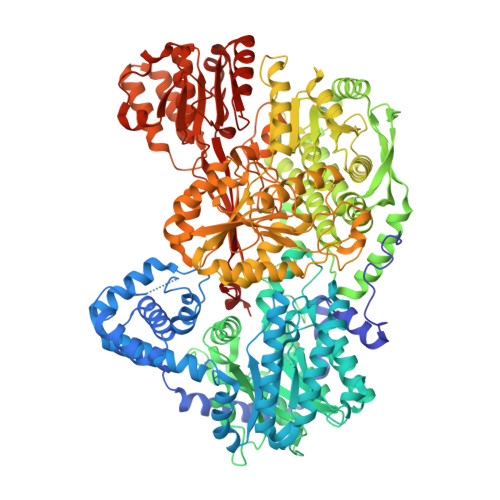
    Proline dehydrogenase (PRODH) is a flavoenzyme that catalyzes the first step of proline catabolism, the oxidation of l-proline to Δ 1 -pyrroline-5-carboxylate. PRODH has emerged as a cancer therapy target because of its involvement in the metabolic reprogramming of cancer cells. Here, we report the discovery of a new class of PRODH inactivator, which covalently and irreversibly modifies the FAD in a light-dependent manner. Two examples, 1,3-dithiolane-2-carboxylate and tetrahydrothiophene-2-carboxylate, have been characterized using X-ray crystallography (1.52-1.85 Å resolution), absorbance spectroscopy, and enzyme kinetics. The structures reveal that in the dark, these compounds function as classical reversible, proline analogue inhibitors. However, exposure of enzyme-inhibitor cocrystals to bright white light induces decarboxylation of the inhibitor and covalent attachment of the residual S-heterocycle to the FAD N5 atom, locking the cofactor into a reduced, inactive state. Spectroscopic measurements of the inactivation process in solution confirm the requirement for light and show that blue light is preferred. Enzyme activity assays show that the rate of inactivation is enhanced by light and that the inactivation is irreversible. We also demonstrate the photosensitivity of cancer cells to one of these compounds. A possible mechanism is proposed involving photoexcitation of the FAD, while the inhibitor is noncovalently bound in the active site, followed by electron transfer, decarboxylation, and radical combination steps. Our results could lead to the development of photopharmacological drugs targeting PRODH.


  • Organizational Affiliation

    Department of Biochemistry, University of Missouri, Columbia, Missouri 65211, United States.


Macromolecules
Find similar proteins by:  (by identity cutoff)  |  3D Structure
Entity ID: 1
MoleculeChains Sequence LengthOrganismDetailsImage
Bifunctional protein PutA
A, B
1,235Sinorhizobium meliloti SM11Mutation(s): 0 
Gene Names: putASM11_chr0102
EC: 1.5.5.2 (PDB Primary Data), 1.2.1.88 (PDB Primary Data)
UniProt
Find proteins for F7X6I3 (Sinorhizobium meliloti (strain SM11))
Explore F7X6I3 
Go to UniProtKB:  F7X6I3
Entity Groups  
Sequence Clusters30% Identity50% Identity70% Identity90% Identity95% Identity100% Identity
UniProt GroupF7X6I3
Sequence Annotations
Expand
  • Reference Sequence
Small Molecules
Ligands 8 Unique
IDChains Name / Formula / InChI Key2D Diagram3D Interactions
FAD (Subject of Investigation/LOI)
Query on FAD

Download Ideal Coordinates CCD File 
F [auth A],
O [auth B]
FLAVIN-ADENINE DINUCLEOTIDE
C27 H33 N9 O15 P2
VWWQXMAJTJZDQX-UYBVJOGSSA-N
NAD (Subject of Investigation/LOI)
Query on NAD

Download Ideal Coordinates CCD File 
G [auth A],
P [auth B]
NICOTINAMIDE-ADENINE-DINUCLEOTIDE
C21 H27 N7 O14 P2
BAWFJGJZGIEFAR-NNYOXOHSSA-N
PGE
Query on PGE

Download Ideal Coordinates CCD File 
E [auth A],
N [auth B]
TRIETHYLENE GLYCOL
C6 H14 O4
ZIBGPFATKBEMQZ-UHFFFAOYSA-N
UJP (Subject of Investigation/LOI)
Query on UJP

Download Ideal Coordinates CCD File 
D [auth A],
M [auth B]
(2R)-thiolane-2-carboxylic acid
C5 H8 O2 S
MZOYMQRKTJRHGJ-SCSAIBSYSA-N
UJM (Subject of Investigation/LOI)
Query on UJM

Download Ideal Coordinates CCD File 
C [auth A],
L [auth B]
(2S)-thiolane-2-carboxylic acid
C5 H8 O2 S
MZOYMQRKTJRHGJ-BYPYZUCNSA-N
PEG
Query on PEG

Download Ideal Coordinates CCD File 
S [auth B]DI(HYDROXYETHYL)ETHER
C4 H10 O3
MTHSVFCYNBDYFN-UHFFFAOYSA-N
SO4
Query on SO4

Download Ideal Coordinates CCD File 
H [auth A],
I [auth A],
J [auth A],
Q [auth B]
SULFATE ION
O4 S
QAOWNCQODCNURD-UHFFFAOYSA-L
MG
Query on MG

Download Ideal Coordinates CCD File 
K [auth A],
R [auth B]
MAGNESIUM ION
Mg
JLVVSXFLKOJNIY-UHFFFAOYSA-N
Experimental Data & Validation

Experimental Data

  • Method: X-RAY DIFFRACTION
  • Resolution: 1.52 Å
  • R-Value Free: 0.186 
  • R-Value Work: 0.164 
  • R-Value Observed: 0.165 
  • Space Group: P 1 21 1
Unit Cell:
Length ( Å )Angle ( ˚ )
a = 101.884α = 90
b = 103.024β = 106.44
c = 127.062γ = 90
Software Package:
Software NamePurpose
PHENIXrefinement
Aimlessdata scaling
PDB_EXTRACTdata extraction
XDSdata reduction
PHENIXphasing

Structure Validation

View Full Validation Report


Ligand Structure Quality Assessment 


Entry History & Funding Information

Deposition Data


Funding OrganizationLocationGrant Number
National Institutes of Health/National Institute of General Medical Sciences (NIH/NIGMS)United StatesR01GM132640

Revision History  (Full details and data files)

  • Version 1.0: 2021-09-29
    Type: Initial release
  • Version 1.1: 2021-12-01
    Changes: Database references
  • Version 1.2: 2023-10-18
    Changes: Data collection, Refinement description