7MTL

Crystal structure of colibactin self-resistance protein ClbS in complex with a dsDNA


Experimental Data Snapshot

  • Method: X-RAY DIFFRACTION
  • Resolution: 2.45 Å
  • R-Value Free: 0.283 
  • R-Value Work: 0.231 
  • R-Value Observed: 0.234 

wwPDB Validation   3D Report Full Report


This is version 1.2 of the entry. See complete history


Literature

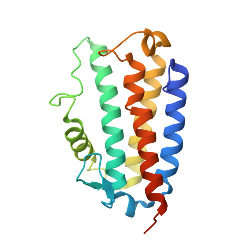
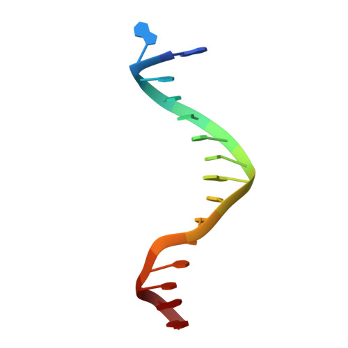
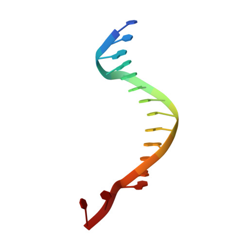
Structural Basis for the Interactions of the Colibactin Resistance Gene Product ClbS with DNA.

Tripathi, P.Bruner, S.D.

(2021) Biochemistry 60: 1619-1625

  • DOI: https://doi.org/10.1021/acs.biochem.1c00201
  • Primary Citation of Related Structures:  
    7MTL, 7MTT

  • PubMed Abstract: 

    The natural product colibactin, along with its associated biosynthetic gene cluster, is an example system for the role microbially derived small molecules play in the human microbiome. This is particularly relevant in the human gut, where host microbiota is involved in various disorders, including colorectal cancer pathogenesis. Bacteria harboring the colibactin gene cluster induce alkylation of nucleobases in host DNA, forming interstrand cross-links both in vivo and in vitro . These lesions can lead to deleterious double-strand breaks and have been identified as the primary mechanism of colibactin-induced cytotoxicity. The gene product ClbS is one of several mechanisms utilized by the producing bacteria to maintain genome integrity. ClbS catalyzes hydrolytic inactivation of colibactin and has been shown to bind DNA, incurring self-resistance. Presented is the molecular basis for ClbS bound to a DNA oligonucleotide. The structure shows the interaction of the protein with the ends of a DNA duplex with terminal nucleotides flipped to the enzyme active site. The structure suggests an additional function for ClbS, the binding to damaged DNA followed by repair. Additionally, our study provides general insight into the function of the widely distributed and largely uncharacterized DUF1706 protein family.


  • Organizational Affiliation

    Department of Chemistry, University of Florida, Gainesville, Florida 32611, United States.


Macromolecules

Find similar proteins by:  (by identity cutoff)  |  3D Structure
Entity ID: 1
MoleculeChains Sequence LengthOrganismDetailsImage
Colibactin self-protection protein ClbS
A, B
178Escherichia coliMutation(s): 0 
Gene Names: 
UniProt
Find proteins for Q0P7K8 (Escherichia coli)
Explore Q0P7K8 
Go to UniProtKB:  Q0P7K8
Entity Groups  
Sequence Clusters30% Identity50% Identity70% Identity90% Identity95% Identity100% Identity
UniProt GroupQ0P7K8
Sequence Annotations
Expand
  • Reference Sequence

Find similar nucleic acids by:  Sequence   |   3D Structure  

Entity ID: 2
MoleculeChains LengthOrganismImage
DNA (5'-D(*AP*AP*TP*TP*CP*CP*CP*AP*CP*TP*TP*CP*CP*AP*G)-3')15synthetic construct
Sequence Annotations
Expand
  • Reference Sequence

Find similar nucleic acids by:  Sequence   |   3D Structure  

Entity ID: 3
MoleculeChains LengthOrganismImage
DNA (5'-D(*CP*TP*GP*GP*AP*AP*GP*TP*GP*GP*GP*AP*AP*TP*T)-3')15synthetic construct
Sequence Annotations
Expand
  • Reference Sequence
Experimental Data & Validation

Experimental Data

  • Method: X-RAY DIFFRACTION
  • Resolution: 2.45 Å
  • R-Value Free: 0.283 
  • R-Value Work: 0.231 
  • R-Value Observed: 0.234 
  • Space Group: P 21 21 21
Unit Cell:
Length ( Å )Angle ( ˚ )
a = 42.783α = 90
b = 75.878β = 90
c = 178.326γ = 90
Software Package:
Software NamePurpose
PHENIXrefinement
PDB_EXTRACTdata extraction
XDSdata reduction
Aimlessdata scaling
PHASERphasing

Structure Validation

View Full Validation Report


Entry History 

Deposition Data

Revision History  (Full details and data files)

  • Version 1.0: 2021-05-26
    Type: Initial release
  • Version 1.1: 2021-06-09
    Changes: Database references
  • Version 1.2: 2023-10-18
    Changes: Data collection, Database references, Refinement description