7MQK

AAC(3)-IIIa in complex with CoA and sisomicin


Experimental Data Snapshot

  • Method: X-RAY DIFFRACTION
  • Resolution: 1.60 Å
  • R-Value Free: 0.204 
  • R-Value Work: 0.179 
  • R-Value Observed: 0.180 

wwPDB Validation   3D Report Full Report


Ligand Structure Quality Assessment 


This is version 1.3 of the entry. See complete history


Literature

Structural elucidation of substrate-bound aminoglycoside acetyltransferase (3)-IIIa.

Zielinski, M.Blanchet, J.Hailemariam, S.Berghuis, A.M.

(2022) PLoS One 17: e0269684-e0269684

  • DOI: https://doi.org/10.1371/journal.pone.0269684
  • Primary Citation of Related Structures:  
    7MQK, 7MQL, 7MQM

  • PubMed Abstract: 

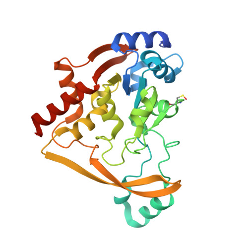
    Canonical aminoglycosides are a large group of antibiotics, where the part of chemical diversity stems from the substitution of the neamine ring system on positions 5 and 6. Certain aminoglycoside modifying enzymes can modify a broad range of 4,5- and 4,6-disubstituted aminoglycosides, with some as many as 15. This study presents the structural and kinetic results describing a promiscuous aminoglycoside acetyltransferase AAC(3)-IIIa. This enzyme has been crystallized in ternary complex with coenzyme A and 4,5- and 4,6-disubstituted aminoglycosides. We have followed up this work with kinetic characterization utilizing a panel of diverse aminoglycosides, including a next-generation aminoglycoside, plazomicin. Lastly, we observed an alternative binding mode of gentamicin in the aminoglycoside binding site, which was proven to be a crystallographic artifact based on mutagenesis.


  • Organizational Affiliation

    Department of Biochemistry, McGill University, Montréal, Québec, Canada.


Macromolecules
Find similar proteins by:  (by identity cutoff)  |  3D Structure
Entity ID: 1
MoleculeChains Sequence LengthOrganismDetailsImage
Aminoglycoside N(3)-acetyltransferase III
A, B, C, D
274Pseudomonas aeruginosaMutation(s): 0 
Gene Names: aacC3
EC: 2.3.1.81
UniProt
Find proteins for P29808 (Pseudomonas aeruginosa)
Explore P29808 
Go to UniProtKB:  P29808
Entity Groups  
Sequence Clusters30% Identity50% Identity70% Identity90% Identity95% Identity100% Identity
UniProt GroupP29808
Sequence Annotations
Expand
  • Reference Sequence
Small Molecules
Ligands 3 Unique
IDChains Name / Formula / InChI Key2D Diagram3D Interactions
COA (Subject of Investigation/LOI)
Query on COA

Download Ideal Coordinates CCD File 
F [auth A],
H [auth B],
K [auth C],
M [auth D]
COENZYME A
C21 H36 N7 O16 P3 S
RGJOEKWQDUBAIZ-IBOSZNHHSA-N
SIS (Subject of Investigation/LOI)
Query on SIS

Download Ideal Coordinates CCD File 
E [auth A],
G [auth B],
J [auth C],
L [auth D]
(1S,2S,3R,4S,6R)-4,6-diamino-3-{[(2S,3R)-3-amino-6-(aminomethyl)-3,4-dihydro-2H-pyran-2-yl]oxy}-2-hydroxycyclohexyl 3-deoxy-4-C-methyl-3-(methylamino)-beta-L-arabinopyranoside
C19 H37 N5 O7
URWAJWIAIPFPJE-YFMIWBNJSA-N
GOL
Query on GOL

Download Ideal Coordinates CCD File 
I [auth B]GLYCEROL
C3 H8 O3
PEDCQBHIVMGVHV-UHFFFAOYSA-N
Modified Residues  1 Unique
IDChains TypeFormula2D DiagramParent
CSO
Query on CSO
A, B, C, D
L-PEPTIDE LINKINGC3 H7 N O3 SCYS
Experimental Data & Validation

Experimental Data

  • Method: X-RAY DIFFRACTION
  • Resolution: 1.60 Å
  • R-Value Free: 0.204 
  • R-Value Work: 0.179 
  • R-Value Observed: 0.180 
  • Space Group: P 21 21 2
Unit Cell:
Length ( Å )Angle ( ˚ )
a = 130.175α = 90
b = 90.927β = 90
c = 100.048γ = 90
Software Package:
Software NamePurpose
PHENIXrefinement
PROTEUM PLUSdata reduction
PROTEUM PLUSdata scaling
PHASERphasing

Structure Validation

View Full Validation Report


Ligand Structure Quality Assessment 


Entry History & Funding Information

Deposition Data


Funding OrganizationLocationGrant Number
Canadian Institutes of Health Research (CIHR)Canada--

Revision History  (Full details and data files)

  • Version 1.0: 2022-07-06
    Type: Initial release
  • Version 1.1: 2022-08-17
    Changes: Database references
  • Version 1.2: 2023-10-18
    Changes: Data collection, Refinement description
  • Version 1.3: 2023-11-15
    Changes: Data collection