


Crystal structure of Candidatus Liberibacter solanacearum dihydrodipicolinate synthase with pyruvate and succinic semi-aldehyde bound in active site
Gilkes, J., Frampton, R.A., Board, A.J., Sheen, C.R., Smith, G.R., Dobson, R.C.J.To be published.
Experimental Data Snapshot
Starting Model: experimental
View more details
Entity ID: 1 | |||||
|---|---|---|---|---|---|
| Molecule | Chains | Sequence Length | Organism | Details | Image |
| 4-hydroxy-tetrahydrodipicolinate synthase | 296 | Candidatus Liberibacter solanacearum | Mutation(s): 0 Gene Names: dapA, DJ66_0589 EC: 4.3.3.7 | 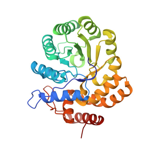 | |
UniProt | |||||
Find proteins for A0A0F4VK59 (Candidatus Liberibacter solanacearum) Explore A0A0F4VK59 Go to UniProtKB: A0A0F4VK59 | |||||
Entity Groups | |||||
| Sequence Clusters | 30% Identity50% Identity70% Identity90% Identity95% Identity100% Identity | ||||
| UniProt Group | A0A0F4VK59 | ||||
Sequence AnnotationsExpand | |||||
Reference Sequence | |||||
| Ligands 2 Unique | |||||
|---|---|---|---|---|---|
| ID | Chains | Name / Formula / InChI Key | 2D Diagram | 3D Interactions | |
| ZGM Download:Ideal Coordinates CCD File | H [auth A] J [auth B] L [auth C] N [auth D] P [auth E] | (4S)-4-hydroxy-2-oxoheptanedioic acid C7 H10 O6 HNOAJOYERZTSNK-BYPYZUCNSA-N |  | ||
| E8U Download:Ideal Coordinates CCD File | G [auth A] I [auth B] K [auth C] M [auth D] O [auth E] | (4R)-4-oxidanyl-2-oxidanylidene-heptanedioic acid C7 H10 O6 HNOAJOYERZTSNK-SCSAIBSYSA-N |  | ||
| Length ( Å ) | Angle ( ˚ ) |
|---|---|
| a = 101.101 | α = 90 |
| b = 132.881 | β = 99.96 |
| c = 155.062 | γ = 90 |
| Software Name | Purpose |
|---|---|
| PHENIX | refinement |
| XDS | data reduction |
| pointless | data scaling |
| PHASER | phasing |


