7MGX

Structure of EmrE-D3 mutant in complex with monobody L10 and methyl viologen


Experimental Data Snapshot

  • Method: X-RAY DIFFRACTION
  • Resolution: 3.13 Å
  • R-Value Free: 0.332 
  • R-Value Work: 0.306 
  • R-Value Observed: 0.307 

wwPDB Validation   3D Report Full Report


Ligand Structure Quality Assessment 


This is version 1.2 of the entry. See complete history


Literature

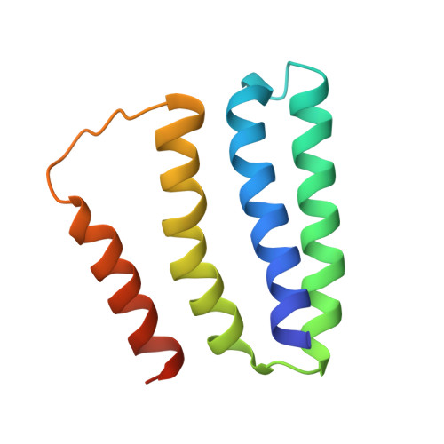
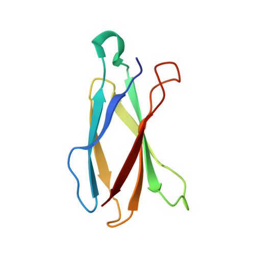
Crystal structures of bacterial small multidrug resistance transporter EmrE in complex with structurally diverse substrates.

Kermani, A.A.Burata, O.E.Koff, B.B.Koide, A.Koide, S.Stockbridge, R.B.

(2022) Elife 11

  • DOI: https://doi.org/10.7554/eLife.76766
  • Primary Citation of Related Structures:  
    7MGX, 7MH6, 7SSU, 7SV9, 7SVX, 7SZT, 7T00

  • PubMed Abstract: 

    Proteins from the bacterial small multidrug resistance (SMR) family are proton-coupled exporters of diverse antiseptics and antimicrobials, including polyaromatic cations and quaternary ammonium compounds. The transport mechanism of the Escherichia coli transporter, EmrE, has been studied extensively, but a lack of high-resolution structural information has impeded a structural description of its molecular mechanism. Here, we apply a novel approach, multipurpose crystallization chaperones, to solve several structures of EmrE, including a 2.9 Å structure at low pH without substrate. We report five additional structures in complex with structurally diverse transported substrates, including quaternary phosphonium, quaternary ammonium, and planar polyaromatic compounds. These structures show that binding site tryptophan and glutamate residues adopt different rotamers to conform to disparate structures without requiring major rearrangements of the backbone structure. Structural and functional comparison to Gdx-Clo, an SMR protein that transports a much narrower spectrum of substrates, suggests that in EmrE, a relatively sparse hydrogen bond network among binding site residues permits increased sidechain flexibility.


  • Organizational Affiliation

    Department of Molecular, Cellular, and Developmental Biology, University of Michigan, Ann Arbor, United States.


Macromolecules
Find similar proteins by:  (by identity cutoff)  |  3D Structure
Entity ID: 1
MoleculeChains Sequence LengthOrganismDetailsImage
Multidrug transporter EmrEA,
C [auth E],
E [auth B],
G [auth F]
110Escherichia coliMutation(s): 3 
Gene Names: emrEebmvrCb0543JW0531
Membrane Entity: Yes 
UniProt
Find proteins for P23895 (Escherichia coli (strain K12))
Explore P23895 
Go to UniProtKB:  P23895
Entity Groups  
Sequence Clusters30% Identity50% Identity70% Identity90% Identity95% Identity100% Identity
UniProt GroupP23895
Sequence Annotations
Expand
  • Reference Sequence
Find similar proteins by:  (by identity cutoff)  |  3D Structure
Entity ID: 2
MoleculeChains Sequence LengthOrganismDetailsImage
L10 MonobodyB [auth C],
D [auth G],
F [auth D],
H
91Homo sapiensMutation(s): 0 
Entity Groups  
Sequence Clusters30% Identity50% Identity70% Identity90% Identity95% Identity100% Identity
Sequence Annotations
Expand
  • Reference Sequence
Experimental Data & Validation

Experimental Data

  • Method: X-RAY DIFFRACTION
  • Resolution: 3.13 Å
  • R-Value Free: 0.332 
  • R-Value Work: 0.306 
  • R-Value Observed: 0.307 
  • Space Group: P 1
Unit Cell:
Length ( Å )Angle ( ˚ )
a = 50.91α = 92.03
b = 75.07β = 90.33
c = 111.43γ = 109.2
Software Package:
Software NamePurpose
PHENIXrefinement
PDB_EXTRACTdata extraction
MOSFLMdata reduction
STARANISOdata scaling
PHASERphasing
Cootmodel building

Structure Validation

View Full Validation Report


Ligand Structure Quality Assessment 


Entry History & Funding Information

Deposition Data


Funding OrganizationLocationGrant Number
National Science Foundation (NSF, United States)United States1845012

Revision History  (Full details and data files)

  • Version 1.0: 2022-03-02
    Type: Initial release
  • Version 1.1: 2022-05-18
    Changes: Database references
  • Version 1.2: 2023-10-18
    Changes: Data collection, Refinement description