A biphenyl inhibitor of eIF4E targeting an internal binding site enables the design of cell-permeable PROTAC-degraders.
Fischer, P.D., Papadopoulos, E., Dempersmier, J.M., Wang, Z.F., Nowak, R.P., Donovan, K.A., Kalabathula, J., Gorgulla, C., Junghanns, P.P.M., Kabha, E., Dimitrakakis, N., Petrov, O.I., Mitsiades, C., Ducho, C., Gelev, V., Fischer, E.S., Wagner, G., Arthanari, H.(2021) Eur J Med Chem 219: 113435-113435
- PubMed: 33892272 Search on PubMedSearch on PubMed Central
- DOI: https://doi.org/10.1016/j.ejmech.2021.113435
- Primary Citation Related Structures:
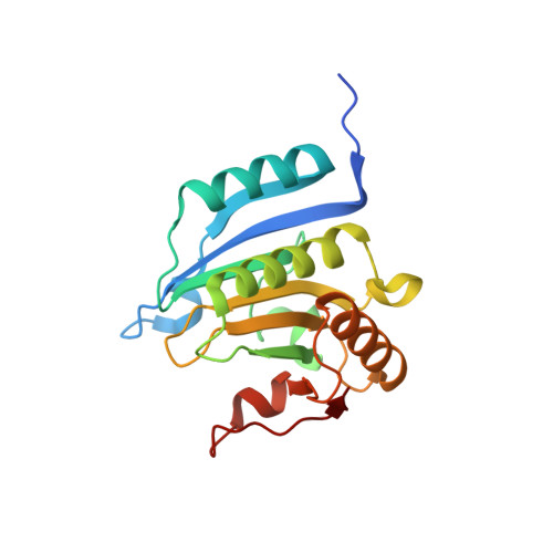
7MEU - PubMed Abstract:
The eukaryotic translation initiation factor 4E (eIF4E) is the master regulator of cap-dependent protein synthesis. Overexpression of eIF4E is implicated in diseases such as cancer, where dysregulation of oncogenic protein translation is frequently observed. eIF4E has been an attractive target for cancer treatment. Here we report a high-resolution X-ray crystal structure of eIF4E in complex with a novel inhibitor (i4EG-BiP) that targets an internal binding site, in contrast to the previously described inhibitor, 4EGI-1, which binds to the surface. We demonstrate that i4EG-BiP is able to displace the scaffold protein eIF4G and inhibit the proliferation of cancer cells. We provide insights into how i4EG-BiP is able to inhibit cap-dependent translation by increasing the eIF4E-4E-BP1 interaction while diminishing the interaction of eIF4E with eIF4G. Leveraging structural details, we designed proteolysis targeted chimeras (PROTACs) derived from 4EGI-1 and i4EG-BiP and characterized these on biochemical and cellular levels. We were able to design PROTACs capable of binding eIF4E and successfully engaging Cereblon, which targets proteins for proteolysis. However, these initial PROTACs did not successfully stimulate degradation of eIF4E, possibly due to competitive effects from 4E-BP1 binding. Our results highlight challenges of targeted proteasomal degradation of eIF4E that must be addressed by future efforts.
- Department of Cancer Biology, Dana-Farber Cancer Institute, Boston, MA, 02215, USA; Department of Biological Chemistry and Molecular Pharmacology, Harvard Medical School, Boston, MA, 02115, USA; Department of Pharmacy, Pharmaceutical and Medicinal Chemistry, Saarland University, Saarbrücken, 66123, Germany.
Organizational Affiliation: