7M8I

Human CYP11B2 and human adrenodoxin in complex with fadrozole


Experimental Data Snapshot

  • Method: X-RAY DIFFRACTION
  • Resolution: 2.94 Å
  • R-Value Free: 0.244 
  • R-Value Work: 0.210 
  • R-Value Observed: 0.211 

wwPDB Validation   3D Report Full Report


Ligand Structure Quality Assessment 


This is version 1.3 of the entry. See complete history


Literature

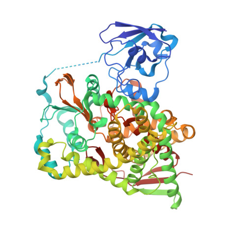
Structural and functional insights into aldosterone synthase interaction with its redox partner protein adrenodoxin.

Brixius-Anderko, S.Scott, E.E.

(2021) J Biol Chem 296: 100794-100794

  • DOI: https://doi.org/10.1016/j.jbc.2021.100794
  • Primary Citation of Related Structures:  
    7M8I

  • PubMed Abstract: 

    Aldosterone is the major mineralocorticoid in the human body controlling blood pressure and salt homeostasis. Overproduction of aldosterone leads to primary aldosteronism, which is the most common form of secondary hypertension with limited treatment options. Production of aldosterone by cytochrome P450 11B2 (CYP11B2, aldosterone synthase) requires two reduction events with the electrons delivered by the iron/sulfur protein adrenodoxin. Very limited information is available about the structural and functional basis of adrenodoxin/CYP11B2 interaction, which impedes the development of new treatment options for primary aldosteronism. A systematic study was carried out to determine if adrenodoxin interaction with CYP11B2 might also have an allosteric component in addition to electron transfer. Indeed, local increases in adrenodoxin concentration promote binding of the substrate 11-deoxycorticosterone and the inhibitor osilodrostat (LCI699) in the active site-over 17 Å away-as well as enhance the inhibitory effect of this latter drug. The CYP11B2 structure in complex with adrenodoxin identified specific residues at the protein-protein interface interacting via five salt bridges and four hydrogen bonds. Comparisons with cholesterol-metabolizing CYP11A1 and cortisol-producing CYP11B1, which also bind adrenodoxin, revealed substantial structural differences in these regions. The structural and functional differences between different P450 interactions with adrenodoxin may provide valuable clues for an orthogonal treatment approach for primary aldosteronism by specifically targeting the interaction between CYP11B2 and adrenodoxin.


  • Organizational Affiliation

    Department of Medicinal Chemistry, University of Michigan, Ann Arbor, Michigan, USA.


Macromolecules
Find similar proteins by:  (by identity cutoff)  |  3D Structure
Entity ID: 1
MoleculeChains Sequence LengthOrganismDetailsImage
Adrenodoxin, Cytochrome P450 11B2, mitochondrial fusion enzyme
A, B, C
609Homo sapiensMutation(s): 0 
Gene Names: FDX1ADXCYP11B2
EC: 1.14.15.5 (PDB Primary Data), 1.14.15.4 (PDB Primary Data)
UniProt & NIH Common Fund Data Resources
Find proteins for P10109 (Homo sapiens)
Explore P10109 
Go to UniProtKB:  P10109
PHAROS:  P10109
GTEx:  ENSG00000137714 
Find proteins for P19099 (Homo sapiens)
Explore P19099 
Go to UniProtKB:  P19099
PHAROS:  P19099
GTEx:  ENSG00000179142 
Entity Groups  
Sequence Clusters30% Identity50% Identity70% Identity90% Identity95% Identity100% Identity
UniProt GroupsP10109P19099
Sequence Annotations
Expand
  • Reference Sequence
Small Molecules
Ligands 3 Unique
IDChains Name / Formula / InChI Key2D Diagram3D Interactions
HEM
Query on HEM

Download Ideal Coordinates CCD File 
E [auth A],
H [auth B],
K [auth C]
PROTOPORPHYRIN IX CONTAINING FE
C34 H32 Fe N4 O4
KABFMIBPWCXCRK-RGGAHWMASA-L
0T3
Query on 0T3

Download Ideal Coordinates CCD File 
F [auth A],
I [auth B],
L [auth C]
4-[(5R)-5,6,7,8-tetrahydroimidazo[1,5-a]pyridin-5-yl]benzonitrile
C14 H13 N3
CLPFFLWZZBQMAO-CQSZACIVSA-N
FES
Query on FES

Download Ideal Coordinates CCD File 
D [auth A],
G [auth B],
J [auth C]
FE2/S2 (INORGANIC) CLUSTER
Fe2 S2
NIXDOXVAJZFRNF-UHFFFAOYSA-N
Experimental Data & Validation

Experimental Data

  • Method: X-RAY DIFFRACTION
  • Resolution: 2.94 Å
  • R-Value Free: 0.244 
  • R-Value Work: 0.210 
  • R-Value Observed: 0.211 
  • Space Group: C 1 2 1
Unit Cell:
Length ( Å )Angle ( ˚ )
a = 140.579α = 90
b = 208.7β = 114.073
c = 124.919γ = 90
Software Package:
Software NamePurpose
PHENIXrefinement
Cootmodel building
HKL-2000data reduction
HKL-2000data scaling
PHASERphasing

Structure Validation

View Full Validation Report


Ligand Structure Quality Assessment 


Entry History & Funding Information

Deposition Data


Funding OrganizationLocationGrant Number
American Heart AssociationUnited States19POST34430199

Revision History  (Full details and data files)

  • Version 1.0: 2021-05-26
    Type: Initial release
  • Version 1.1: 2021-06-02
    Changes: Database references
  • Version 1.2: 2021-07-14
    Changes: Database references
  • Version 1.3: 2023-10-18
    Changes: Data collection, Database references, Refinement description