7M1T | pdb_00007m1t

Crystal structure of an archaeal CNNM, MtCorB, with C-terminal deletion in complex with Mg2+-ATP


Experimental Data Snapshot

  • Method: X-RAY DIFFRACTION
  • Resolution: 3.26 Å
  • R-Value Free: 
    0.267 (Depositor), 0.267 (DCC) 
  • R-Value Work: 
    0.226 (Depositor), 0.228 (DCC) 
  • R-Value Observed: 
    0.228 (Depositor) 

Starting Model: experimental
View more details

wwPDB Validation 3D Report Full Report

Validation slider image for 7M1T

Ligand Structure Quality Assessment 


This is version 1.2 of the entry. See complete history

Literature

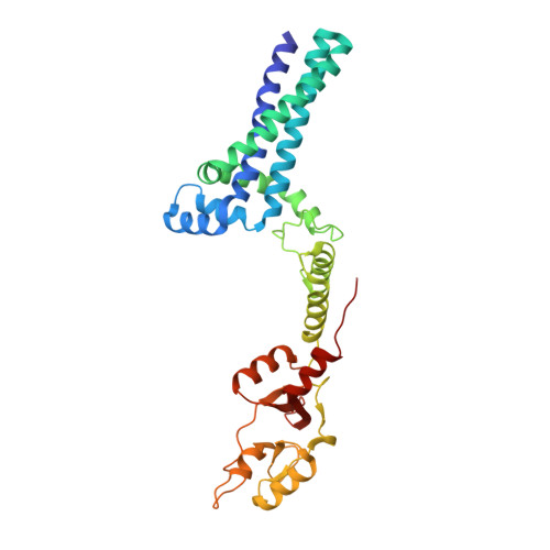
Crystal structure of an archaeal CorB magnesium transporter.

Chen, Y.S.Kozlov, G.Moeller, B.E.Rohaim, A.Fakih, R.Roux, B.Burke, J.E.Gehring, K.

(2021) Nat Commun 12: 4028-4028

  • DOI: https://doi.org/10.1038/s41467-021-24282-7
  • Primary Citation Related Structures: 
    7M1T, 7M1U, 7MSU

  • PubMed Abstract: 

    CNNM/CorB proteins are a broadly conserved family of integral membrane proteins with close to 90,000 protein sequences known. They are associated with Mg 2+ transport but it is not known if they mediate transport themselves or regulate other transporters. Here, we determine the crystal structure of an archaeal CorB protein in two conformations (apo and Mg 2+ -ATP bound). The transmembrane DUF21 domain exists in an inward-facing conformation with a Mg 2+ ion coordinated by a conserved π-helix. In the absence of Mg 2+ -ATP, the CBS-pair domain adopts an elongated dimeric configuration with previously unobserved domain-domain contacts. Hydrogen-deuterium exchange mass spectrometry, analytical ultracentrifugation, and molecular dynamics experiments support a role of the structural rearrangements in mediating Mg 2+ -ATP sensing. Lastly, we use an in vitro, liposome-based assay to demonstrate direct Mg 2+ transport by CorB proteins. These structural and functional insights provide a framework for understanding function of CNNMs in Mg 2+ transport and associated diseases.


  • Organizational Affiliation
    • Department of Biochemistry & Centre de Recherche en Biologie Structurale, McGill University, Montréal, QC, Canada.

Macromolecule Content 

  • Total Structure Weight: 79.18 kDa 
  • Atom Count: 5,085 
  • Modeled Residue Count: 637 
  • Deposited Residue Count: 652 
  • Unique protein chains: 1

Macromolecules

Find similar proteins by:|  3D Structure
Entity ID: 1
MoleculeChains  Sequence LengthOrganismDetailsImage
Hemolysin, contains CBS domains
A, B
326Methanoculleus thermophilusMutation(s): 0 
Gene Names: SAMN04488571_101329
Membrane Entity: Yes 
UniProt
Find proteins for A0A1G8XA46 (Methanoculleus thermophilus)
Explore A0A1G8XA46 
Go to UniProtKB:  A0A1G8XA46
Entity Groups
Sequence Clusters30% Identity50% Identity70% Identity90% Identity95% Identity100% Identity
UniProt GroupA0A1G8XA46
Sequence Annotations
Expand
Reference Sequence

Small Molecules

Ligands 5 Unique
IDChains Name / Formula / InChI Key2D Diagram3D Interactions
ATP

Query on ATP


Download:Ideal Coordinates CCD File
J [auth A],
T [auth B]
ADENOSINE-5'-TRIPHOSPHATE
C10 H16 N5 O13 P3
ZKHQWZAMYRWXGA-KQYNXXCUSA-N
UMQ

Query on UMQ


Download:Ideal Coordinates CCD File
E [auth A]
F [auth A]
G [auth A]
H [auth A]
I [auth A]
E [auth A],
F [auth A],
G [auth A],
H [auth A],
I [auth A],
O [auth B],
P [auth B],
Q [auth B],
R [auth B],
S [auth B]
UNDECYL-MALTOSIDE
C23 H44 O11
UYEMNFYVTFDKRG-ZNGNCRBCSA-N
1PE

Query on 1PE


Download:Ideal Coordinates CCD File
K [auth A]PENTAETHYLENE GLYCOL
C10 H22 O6
JLFNLZLINWHATN-UHFFFAOYSA-N
SO4

Query on SO4


Download:Ideal Coordinates CCD File
D [auth A],
N [auth B]
SULFATE ION
O4 S
QAOWNCQODCNURD-UHFFFAOYSA-L
MG

Query on MG


Download:Ideal Coordinates CCD File
C [auth A],
L [auth B],
M [auth B]
MAGNESIUM ION
Mg
JLVVSXFLKOJNIY-UHFFFAOYSA-N

Experimental Data & Validation

Experimental Data

  • Method: X-RAY DIFFRACTION
  • Resolution: 3.26 Å
  • R-Value Free:  0.267 (Depositor), 0.267 (DCC) 
  • R-Value Work:  0.226 (Depositor), 0.228 (DCC) 
  • R-Value Observed: 0.228 (Depositor) 
Space Group: P 21 21 21
Unit Cell:
Length ( Å )Angle ( ˚ )
a = 61.05α = 90
b = 118.676β = 90
c = 177.305γ = 90
Software Package:
Software NamePurpose
HKL-2000data reduction
HKL-2000data scaling
PHENIXphasing
PHENIXrefinement

Structure Validation

View Full Validation Report


Ligand Structure Quality Assessment 


Entry History 

& Funding Information

Deposition Data


Funding OrganizationLocationGrant Number
Natural Sciences and Engineering Research Council (NSERC, Canada)CanadaRGPIN-2020-07195

Revision History  (Full details and data files)

  • Version 1.0: 2021-06-16
    Type: Initial release
  • Version 1.1: 2021-07-07
    Changes: Database references
  • Version 1.2: 2023-10-18
    Changes: Data collection, Database references, Refinement description