7M18

HeLa-tubulin in complex with cryptophycin 1

  • Classification: CELL CYCLE
  • Organism(s): Homo sapiens
  • Mutation(s): No 

  • Deposited: 2021-03-12 Released: 2021-09-08 
  • Deposition Author(s): Eren, E.
  • Funding Organization(s): National Institutes of Health/National Institute of Arthritis and Musculoskeletal and Skin Diseases (NIH/NIAMS)

Experimental Data Snapshot

  • Method: ELECTRON MICROSCOPY
  • Resolution: 3.38 Å
  • Aggregation State: PARTICLE 
  • Reconstruction Method: SINGLE PARTICLE 

wwPDB Validation   3D Report Full Report


This is version 1.2 of the entry. See complete history


Literature

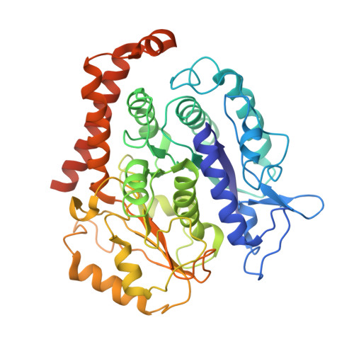
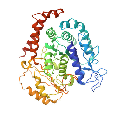
Conformational changes in tubulin upon binding cryptophycin-52 reveal its mechanism of action.

Eren, E.Watts, N.R.Sackett, D.L.Wingfield, P.T.

(2021) J Biol Chem 297: 101138-101138

  • DOI: https://doi.org/10.1016/j.jbc.2021.101138
  • Primary Citation of Related Structures:  
    7LXB, 7M18, 7M20

  • PubMed Abstract: 

    Cryptophycin-52 (Cp-52) is potentially the most potent anticancer drug known, with IC 50 values in the low picomolar range, but its binding site on tubulin and mechanism of action are unknown. Here, we have determined the binding site of Cp-52, and its parent compound, cryptophycin-1, on HeLa tubulin, to a resolution of 3.3 Å and 3.4 Å, respectively, by cryo-EM and characterized this binding further by molecular dynamics simulations. The binding site was determined to be located at the tubulin interdimer interface and partially overlap that of maytansine, another cytotoxic tubulin inhibitor. Binding induces curvature both within and between tubulin dimers that is incompatible with the microtubule lattice. Conformational changes occur in both α-tubulin and β-tubulin, particularly in helices H8 and H10, with distinct differences between α and β monomers and between Cp-52-bound and cryptophycin-1-bound tubulin. From these results, we have determined: (i) the mechanism of action of inhibition of both microtubule polymerization and depolymerization, (ii) how the affinity of Cp-52 for tubulin may be enhanced, and (iii) where linkers for targeted delivery can be optimally attached to this molecule.


  • Organizational Affiliation

    Protein Expression Laboratory, NIAMS, National Institutes of Health, Bethesda, Maryland, USA.


Macromolecules
Find similar proteins by:  (by identity cutoff)  |  3D Structure
Entity ID: 1
MoleculeChains Sequence LengthOrganismDetailsImage
Tubulin beta-3 chain450Homo sapiensMutation(s): 0 
UniProt & NIH Common Fund Data Resources
Find proteins for Q13509 (Homo sapiens)
Explore Q13509 
Go to UniProtKB:  Q13509
PHAROS:  Q13509
GTEx:  ENSG00000258947 
Entity Groups  
Sequence Clusters30% Identity50% Identity70% Identity90% Identity95% Identity100% Identity
UniProt GroupQ13509
Sequence Annotations
Expand
  • Reference Sequence
Find similar proteins by:  (by identity cutoff)  |  3D Structure
Entity ID: 2
MoleculeChains Sequence LengthOrganismDetailsImage
Tubulin alpha-1B chain451Homo sapiensMutation(s): 0 
UniProt & NIH Common Fund Data Resources
Find proteins for P68363 (Homo sapiens)
Explore P68363 
Go to UniProtKB:  P68363
PHAROS:  P68363
GTEx:  ENSG00000123416 
Entity Groups  
Sequence Clusters30% Identity50% Identity70% Identity90% Identity95% Identity100% Identity
UniProt GroupP68363
Sequence Annotations
Expand
  • Reference Sequence
Small Molecules
Ligands 3 Unique
IDChains Name / Formula / InChI Key2D Diagram3D Interactions
YNP
Query on YNP

Download Ideal Coordinates CCD File 
CA [auth J]
FA [auth L]
IA [auth N]
LA [auth P]
Q [auth B]
CA [auth J],
FA [auth L],
IA [auth N],
LA [auth P],
Q [auth B],
T [auth D],
W [auth F],
Z [auth H]
Cryptophycin 1
C35 H43 Cl N2 O8
PSNOPSMXOBPNNV-XUMGCJJFSA-N
GTP
Query on GTP

Download Ideal Coordinates CCD File 
BA [auth G]
EA [auth I]
HA [auth K]
KA [auth M]
NA [auth O]
BA [auth G],
EA [auth I],
HA [auth K],
KA [auth M],
NA [auth O],
S [auth A],
V [auth C],
Y [auth E]
GUANOSINE-5'-TRIPHOSPHATE
C10 H16 N5 O14 P3
XKMLYUALXHKNFT-UUOKFMHZSA-N
GDP
Query on GDP

Download Ideal Coordinates CCD File 
AA [auth H]
DA [auth J]
GA [auth L]
JA [auth N]
MA [auth P]
AA [auth H],
DA [auth J],
GA [auth L],
JA [auth N],
MA [auth P],
R [auth B],
U [auth D],
X [auth F]
GUANOSINE-5'-DIPHOSPHATE
C10 H15 N5 O11 P2
QGWNDRXFNXRZMB-UUOKFMHZSA-N
Experimental Data & Validation

Experimental Data

  • Method: ELECTRON MICROSCOPY
  • Resolution: 3.38 Å
  • Aggregation State: PARTICLE 
  • Reconstruction Method: SINGLE PARTICLE 

Structure Validation

View Full Validation Report


Entry History & Funding Information

Deposition Data

  • Released Date: 2021-09-08 
  • Deposition Author(s): Eren, E.

Funding OrganizationLocationGrant Number
National Institutes of Health/National Institute of Arthritis and Musculoskeletal and Skin Diseases (NIH/NIAMS)United StatesIntramural Research

Revision History  (Full details and data files)

  • Version 1.0: 2021-09-08
    Type: Initial release
  • Version 1.1: 2021-09-15
    Changes: Database references
  • Version 1.2: 2021-10-06
    Changes: Database references