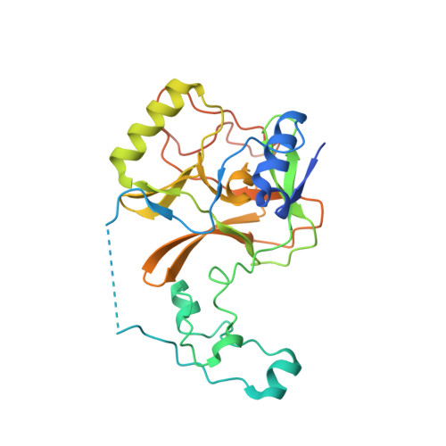
7LZF

Crystal Structure of SETD2 bound to Compound 57


Experimental Data Snapshot

  • Method: X-RAY DIFFRACTION
  • Resolution: 2.47 Å
  • R-Value Free: 0.263 
  • R-Value Work: 0.216 
  • R-Value Observed: 0.218 

wwPDB Validation   3D Report Full Report


Ligand Structure Quality Assessment 


This is version 1.2 of the entry. See complete history


Literature

Discovery of a First-in-Class Inhibitor of the Histone Methyltransferase SETD2 Suitable for Preclinical Studies.

Lampe, J.W.Alford, J.S.Boriak-Sjodin, P.A.Brach, D.Cosmopoulos, K.Duncan, K.W.Eckley, S.T.Foley, M.A.Harvey, D.M.Motwani, V.Munchhof, M.J.Raimondi, A.Riera, T.V.Tang, C.Thomenius, M.J.Totman, J.Farrow, N.A.

(2021) ACS Med Chem Lett 12: 1539-1545

  • DOI: https://doi.org/10.1021/acsmedchemlett.1c00272
  • Primary Citation of Related Structures:  
    7LZB, 7LZD, 7LZF

  • PubMed Abstract: 

    SET domain-containing protein 2 (SETD2), a histone methyltransferase, has been identified as a target of interest in certain hematological malignancies, including multiple myeloma. This account details the discovery of EPZ-719 , a novel and potent SETD2 inhibitor with a high selectivity over other histone methyltransferases. A screening campaign of the Epizyme proprietary histone methyltransferase-biased library identified potential leads based on a 2-amidoindole core. Structure-based drug design (SBDD) and drug metabolism/pharmacokinetics (DMPK) optimization resulted in EPZ-719 , an attractive tool compound for the interrogation of SETD2 biology that enables in vivo target validation studies.


  • Organizational Affiliation

    Epizyme Inc., 400 Technology Square, Fourth Floor, Cambridge, Massachusetts 02139, United States.


Macromolecules
Find similar proteins by:  (by identity cutoff)  |  3D Structure
Entity ID: 1
MoleculeChains Sequence LengthOrganismDetailsImage
Histone-lysine N-methyltransferase SETD2278Homo sapiensMutation(s): 0 
Gene Names: SETD2HIF1HYPBKIAA1732KMT3ASET2HSPC069
EC: 2.1.1.359 (PDB Primary Data), 2.1.1 (PDB Primary Data)
UniProt & NIH Common Fund Data Resources
Find proteins for Q9BYW2 (Homo sapiens)
Explore Q9BYW2 
Go to UniProtKB:  Q9BYW2
PHAROS:  Q9BYW2
GTEx:  ENSG00000181555 
Entity Groups  
Sequence Clusters30% Identity50% Identity70% Identity90% Identity95% Identity100% Identity
UniProt GroupQ9BYW2
Sequence Annotations
Expand
  • Reference Sequence
Small Molecules
Ligands 4 Unique
IDChains Name / Formula / InChI Key2D Diagram3D Interactions
YHV (Subject of Investigation/LOI)
Query on YHV

Download Ideal Coordinates CCD File 
F [auth A]4-fluoro-N-[(1R,3S)-3-{(3S)-3-[(methanesulfonyl)(methyl)amino]pyrrolidin-1-yl}cyclohexyl]-7-methyl-1H-indole-2-carboxamide
C22 H31 F N4 O3 S
ZLVKIBLQXFILMA-IKGGRYGDSA-N
SAM
Query on SAM

Download Ideal Coordinates CCD File 
E [auth A]S-ADENOSYLMETHIONINE
C15 H22 N6 O5 S
MEFKEPWMEQBLKI-FCKMPRQPSA-N
ZN
Query on ZN

Download Ideal Coordinates CCD File 
B [auth A],
C [auth A],
D [auth A]
ZINC ION
Zn
PTFCDOFLOPIGGS-UHFFFAOYSA-N
EDO
Query on EDO

Download Ideal Coordinates CCD File 
G [auth A]1,2-ETHANEDIOL
C2 H6 O2
LYCAIKOWRPUZTN-UHFFFAOYSA-N
Experimental Data & Validation

Experimental Data

  • Method: X-RAY DIFFRACTION
  • Resolution: 2.47 Å
  • R-Value Free: 0.263 
  • R-Value Work: 0.216 
  • R-Value Observed: 0.218 
  • Space Group: P 21 21 21
Unit Cell:
Length ( Å )Angle ( ˚ )
a = 49.91α = 90
b = 76.648β = 90
c = 76.186γ = 90
Software Package:
Software NamePurpose
REFMACrefinement
PDB_EXTRACTdata extraction
XDSdata reduction
Aimlessdata scaling

Structure Validation

View Full Validation Report


Ligand Structure Quality Assessment 


Entry History 

Deposition Data

Revision History  (Full details and data files)

  • Version 1.0: 2021-09-22
    Type: Initial release
  • Version 1.1: 2021-11-03
    Changes: Database references
  • Version 1.2: 2023-10-18
    Changes: Data collection, Refinement description