7LUO

N-terminus of Skp2 bound to Cyclin A


Experimental Data Snapshot

  • Method: X-RAY DIFFRACTION
  • Resolution: 3.17 Å
  • R-Value Free: 0.277 
  • R-Value Work: 0.234 
  • R-Value Observed: 0.236 

wwPDB Validation   3D Report Full Report


This is version 1.3 of the entry. See complete history


Literature

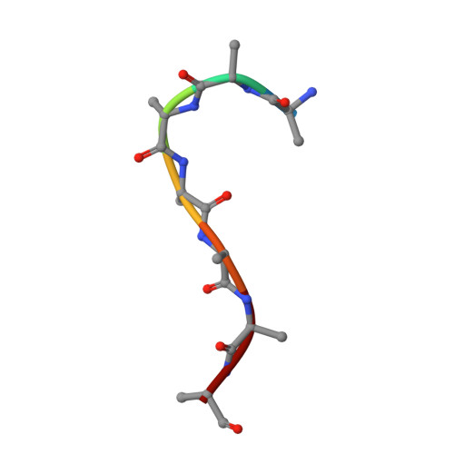
Bipartite binding of the N terminus of Skp2 to cyclin A.

Kelso, S.Orlicky, S.Beenstock, J.Ceccarelli, D.F.Kurinov, I.Gish, G.Sicheri, F.

(2021) Structure 29: 975

  • DOI: https://doi.org/10.1016/j.str.2021.04.011
  • Primary Citation of Related Structures:  
    7LUO

  • PubMed Abstract: 

    Skp2 and cyclin A are cell-cycle regulators that control the activity of CDK2. Cyclin A acts as an activator and substrate recruitment factor of CDK2, while Skp2 mediates the ubiquitination and subsequent destruction of the CDK inhibitor protein p27. The N terminus of Skp2 can interact directly with cyclin A but is not required for p27 ubiquitination. To gain insight into this poorly understood interaction, we have solved the 3.2 Å X-ray crystal structure of the N terminus of Skp2 bound to cyclin A. The structure reveals a bipartite mode of interaction with two motifs in Skp2 recognizing two discrete surfaces on cyclin A. The uncovered binding mechanism allows for a rationalization of the inhibitory effect of Skp2 on CDK2-cyclin A kinase activity toward the RxL motif containing substrates and raises the possibility that other intermolecular regulators and substrates may use similar non-canonical modes of interaction for cyclin targeting.


  • Organizational Affiliation

    Lunenfeld-Tanenbaum Research Institute, Sinai Health System, Toronto, ON M5G 1X5, Canada; Department of Molecular Genetics, University of Toronto, ON M5S 1A8, Canada.


Macromolecules
Find similar proteins by:  (by identity cutoff)  |  3D Structure
Entity ID: 1
MoleculeChains Sequence LengthOrganismDetailsImage
S-phase kinase-associated protein 2,Cyclin-A2
A, C
335Homo sapiensMutation(s): 0 
Gene Names: SKP2FBXL1CCNA2CCN1CCNA
UniProt & NIH Common Fund Data Resources
Find proteins for P20248 (Homo sapiens)
Explore P20248 
Go to UniProtKB:  P20248
PHAROS:  P20248
GTEx:  ENSG00000145386 
Find proteins for Q13309 (Homo sapiens)
Explore Q13309 
Go to UniProtKB:  Q13309
PHAROS:  Q13309
GTEx:  ENSG00000145604 
Entity Groups  
Sequence Clusters30% Identity50% Identity70% Identity90% Identity95% Identity100% Identity
UniProt GroupsQ13309P20248
Sequence Annotations
Expand
  • Reference Sequence

Find similar proteins by:  Sequence   |   3D Structure  

Entity ID: 2
MoleculeChains Sequence LengthOrganismDetailsImage
Skp2 Motif 1 uncharacterized fragment 18Homo sapiensMutation(s): 0 
Sequence Annotations
Expand
  • Reference Sequence

Find similar proteins by:  Sequence   |   3D Structure  

Entity ID: 3
MoleculeChains Sequence LengthOrganismDetailsImage
Skp2 Motif 1 uncharacterized fragment 27Homo sapiensMutation(s): 0 
Sequence Annotations
Expand
  • Reference Sequence
Experimental Data & Validation

Experimental Data

  • Method: X-RAY DIFFRACTION
  • Resolution: 3.17 Å
  • R-Value Free: 0.277 
  • R-Value Work: 0.234 
  • R-Value Observed: 0.236 
  • Space Group: P 4 21 2
Unit Cell:
Length ( Å )Angle ( ˚ )
a = 109.738α = 90
b = 109.738β = 90
c = 152.03γ = 90
Software Package:
Software NamePurpose
PHENIXrefinement
XDSdata reduction
Aimlessdata scaling
PDB_EXTRACTdata extraction
PHASERphasing

Structure Validation

View Full Validation Report


Entry History & Funding Information

Deposition Data


Funding OrganizationLocationGrant Number
Canadian Institutes of Health Research (CIHR)CanadaFDN-143277

Revision History  (Full details and data files)

  • Version 1.0: 2021-05-12
    Type: Initial release
  • Version 1.1: 2021-05-26
    Changes: Database references
  • Version 1.2: 2021-09-15
    Changes: Database references
  • Version 1.3: 2023-10-18
    Changes: Data collection, Refinement description