7LRX

Structure of HIV-1 Reverse Transcriptase in complex with DNA, L-dCTP, and CA(2+) ion


Experimental Data Snapshot

  • Method: X-RAY DIFFRACTION
  • Resolution: 2.90 Å
  • R-Value Free: 0.247 
  • R-Value Work: 0.208 
  • R-Value Observed: 0.209 

wwPDB Validation   3D Report Full Report


Ligand Structure Quality Assessment 


This is version 1.3 of the entry. See complete history


Literature

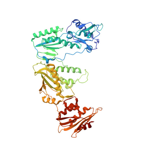
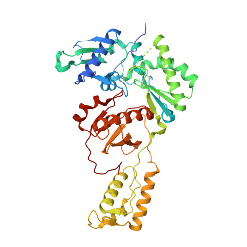
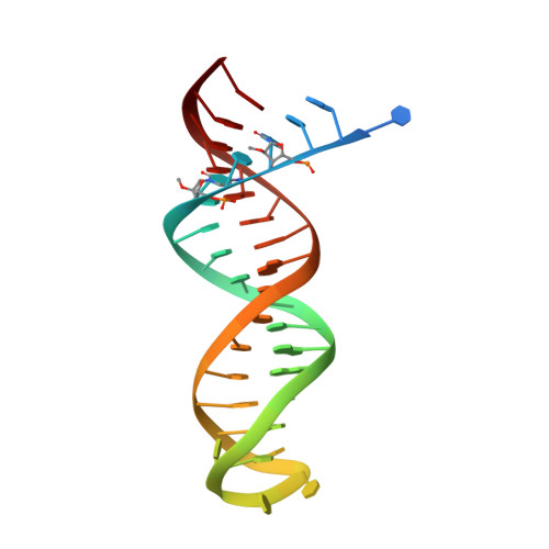
Structural basis of HIV inhibition by L-nucleosides: Opportunities for drug development and repurposing.

Ruiz, F.X.Hoang, A.Dilmore, C.R.DeStefano, J.J.Arnold, E.

(2022) Drug Discov Today 27: 1832-1846

  • DOI: https://doi.org/10.1016/j.drudis.2022.02.016
  • Primary Citation of Related Structures:  
    7LRI, 7LRM, 7LRX, 7LRY, 7LSK

  • PubMed Abstract: 

    Infection with HIV can cripple the immune system and lead to AIDS. Hepatitis B virus (HBV) is a hepadnavirus that causes human liver diseases. Both pathogens are major public health problems affecting millions of people worldwide. The polymerases from both viruses are the most common drug target for viral inhibition, sharing common architecture at their active sites. The L-nucleoside drugs emtricitabine and lamivudine are widely used HIV reverse transcriptase (RT) and HBV polymerase (Pol) inhibitors. Nevertheless, structural details of their binding to RT(Pol)/nucleic acid remained unknown until recently. Here, we discuss the implications of these structures, alongside related complexes with L-dNTPs, for the development of novel L-nucleos(t)ide drugs, and prospects for repurposing them.


  • Organizational Affiliation

    Center for Advanced Biotechnology and Medicine, and Department of Chemistry and Chemical Biology, Rutgers University, Piscataway, NJ 08854, USA. Electronic address: xavier@cabm.rutgers.edu.


Macromolecules

Find similar proteins by:  (by identity cutoff)  |  3D Structure
Entity ID: 1
MoleculeChains Sequence LengthOrganismDetailsImage
Reverse transcriptase p66
A, C
555Human immunodeficiency virus 1Mutation(s): 2 
EC: 2.7.7.49 (PDB Primary Data), 2.7.7.7 (PDB Primary Data), 3.1.26.13 (PDB Primary Data)
UniProt
Find proteins for P03366 (Human immunodeficiency virus type 1 group M subtype B (isolate BH10))
Explore P03366 
Go to UniProtKB:  P03366
Entity Groups  
Sequence Clusters30% Identity50% Identity70% Identity90% Identity95% Identity100% Identity
UniProt GroupP03366
Sequence Annotations
Expand
  • Reference Sequence
Find similar proteins by:  (by identity cutoff)  |  3D Structure
Entity ID: 2
MoleculeChains Sequence LengthOrganismDetailsImage
Reverse transcriptase p51
B, D
429Human immunodeficiency virus 1Mutation(s): 1 
EC: 2.7.7.49
UniProt
Find proteins for P03366 (Human immunodeficiency virus type 1 group M subtype B (isolate BH10))
Explore P03366 
Go to UniProtKB:  P03366
Entity Groups  
Sequence Clusters30% Identity50% Identity70% Identity90% Identity95% Identity100% Identity
UniProt GroupP03366
Sequence Annotations
Expand
  • Reference Sequence
Find similar nucleic acids by:  (by identity cutoff)  |  3D Structure
Entity ID: 3
MoleculeChains LengthOrganismImage
DNA/RNA (38-MER)E [auth F],
F [auth E]
38synthetic construct
Sequence Annotations
Expand
  • Reference Sequence
Oligosaccharides

Help

Entity ID: 4
MoleculeChains Length2D Diagram Glycosylation3D Interactions
beta-D-fructofuranose-(2-1)-alpha-D-glucopyranoseG [auth H],
H [auth L]
2N/A
Glycosylation Resources
GlyTouCan:  G05551OP
GlyCosmos:  G05551OP
Small Molecules
Ligands 5 Unique
IDChains Name / Formula / InChI Key2D Diagram3D Interactions
1S0 (Subject of Investigation/LOI)
Query on 1S0

Download Ideal Coordinates CCD File 
J [auth A],
O [auth C]
4-amino-1-{2-deoxy-5-O-[(R)-hydroxy{[(S)-hydroxy(phosphonooxy)phosphoryl]oxy}phosphoryl]-beta-L-erythro-pentofuranosyl}pyrimidin-2(1H)-one
C9 H16 N3 O13 P3
RGWHQCVHVJXOKC-CHKWXVPMSA-N
SO4
Query on SO4

Download Ideal Coordinates CCD File 
I [auth A],
N [auth C]
SULFATE ION
O4 S
QAOWNCQODCNURD-UHFFFAOYSA-L
GOL
Query on GOL

Download Ideal Coordinates CCD File 
M [auth B],
R [auth D]
GLYCEROL
C3 H8 O3
PEDCQBHIVMGVHV-UHFFFAOYSA-N
CA
Query on CA

Download Ideal Coordinates CCD File 
K [auth A],
P [auth C]
CALCIUM ION
Ca
BHPQYMZQTOCNFJ-UHFFFAOYSA-N
NH4
Query on NH4

Download Ideal Coordinates CCD File 
L [auth A],
Q [auth C]
AMMONIUM ION
H4 N
QGZKDVFQNNGYKY-UHFFFAOYSA-O
Experimental Data & Validation

Experimental Data

  • Method: X-RAY DIFFRACTION
  • Resolution: 2.90 Å
  • R-Value Free: 0.247 
  • R-Value Work: 0.208 
  • R-Value Observed: 0.209 
  • Space Group: P 1 21 1
Unit Cell:
Length ( Å )Angle ( ˚ )
a = 89.881α = 90
b = 132.622β = 97.763
c = 139.376γ = 90
Software Package:
Software NamePurpose
PHENIXrefinement
MOSFLMdata reduction
Aimlessdata scaling
PHASERphasing

Structure Validation

View Full Validation Report


Ligand Structure Quality Assessment 


Entry History & Funding Information

Deposition Data


Funding OrganizationLocationGrant Number
National Institutes of Health/National Institute Of Allergy and Infectious Diseases (NIH/NIAID)United StatesR37 AI027690

Revision History  (Full details and data files)

  • Version 1.0: 2022-02-23
    Type: Initial release
  • Version 1.1: 2022-03-09
    Changes: Database references
  • Version 1.2: 2022-07-06
    Changes: Database references
  • Version 1.3: 2023-10-18
    Changes: Data collection, Refinement description