Discovery of Potent Coumarin-Based Kinetic Stabilizers of Amyloidogenic Immunoglobulin Light Chains Using Structure-Based Design.
Yan, N.L., Santos-Martins, D., Nair, R., Chu, A., Wilson, I.A., Johnson, K.A., Forli, S., Morgan, G.J., Petrassi, H.M., Kelly, J.W.(2021) J Med Chem 64: 6273-6299
- PubMed: 33939422 Search on PubMedSearch on PubMed Central
- DOI: https://doi.org/10.1021/acs.jmedchem.1c00339
- Primary Citation Related Structures:
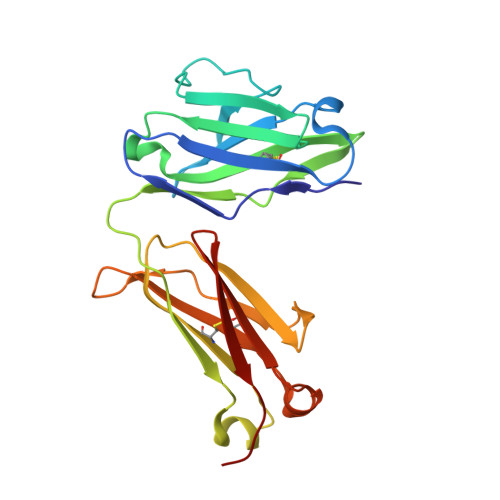
7LMN, 7LMO, 7LMP, 7LMQ, 7LMR - PubMed Abstract:
In immunoglobulin light-chain (LC) amyloidosis, transient unfolding or unfolding and proteolysis enable aggregation of LC proteins, causing potentially fatal organ damage. A drug that kinetically stabilizes LCs could suppress aggregation; however, LC sequences are variable and have no natural ligands, hindering drug development efforts. We previously identified high-throughput screening hits that bind to a site at the interface between the two variable domains of the LC homodimer. We hypothesized that extending the stabilizers beyond this initially characterized binding site would improve affinity. Here, using protease sensitivity assays, we identified stabilizers that can be divided into four substructures. Some stabilizers exhibit nanomolar EC 50 values, a 3000-fold enhancement over the screening hits. Crystal structures reveal a key π-π stacking interaction with a conserved tyrosine residue that was not utilized by the screening hits. These data provide a foundation for developing LC stabilizers with improved binding selectivity and enhanced physicochemical properties.
- Department of Chemistry, The Scripps Research Institute, La Jolla, California 92037, United States.
Organizational Affiliation: