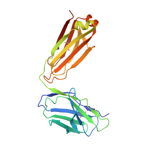
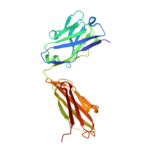
7LJA

Human TRAAK K+ channel FHEIG mutant A198E in a Tl+ bound conductive conformation


Experimental Data Snapshot

  • Method: X-RAY DIFFRACTION
  • Resolution: 2.77 Å
  • R-Value Free: 0.243 
  • R-Value Work: 0.201 
  • R-Value Observed: 0.203 

wwPDB Validation   3D Report Full Report


This is version 1.3 of the entry. See complete history


Literature

Physical basis for distinct basal and mechanically gated activity of the human K + channel TRAAK.

Rietmeijer, R.A.Sorum, B.Li, B.Brohawn, S.G.

(2021) Neuron 109: 2902-2913.e4

  • DOI: https://doi.org/10.1016/j.neuron.2021.07.009
  • Primary Citation of Related Structures:  
    7LJ4, 7LJ5, 7LJA, 7LJB

  • PubMed Abstract: 

    TRAAK is a mechanosensitive two-pore domain K + (K2P) channel localized to nodes of Ranvier in myelinated neurons. TRAAK deletion in mice results in mechanical and thermal allodynia, and gain-of-function mutations cause the human neurodevelopmental disorder FHEIG. TRAAK displays basal and stimulus-gated activities typical of K2Ps, but the mechanistic and structural differences between these modes are unknown. Here, we demonstrate that basal and mechanically gated openings are distinguished by their conductance, kinetics, and structure. Basal openings are low conductance, short duration, and due to a conductive channel conformation with the interior cavity exposed to the surrounding membrane. Mechanically gated openings are high conductance, long duration, and due to a channel conformation in which the interior cavity is sealed to the surrounding membrane. Our results explain how dual modes of activity are produced by a single ion channel and provide a basis for the development of state-selective pharmacology with the potential to treat disease.


  • Organizational Affiliation

    Department of Molecular & Cell Biology, University of California Berkeley, Berkeley, CA 94720, USA; Helen Wills Neuroscience Institute, University of California Berkeley, Berkeley, CA 94720, USA; Biophysics Graduate Program, University of California Berkeley, Berkeley, CA 94720, USA; California Institute for Quantitative Biology (QB3), University of California Berkeley, Berkeley, CA 94720, USA.


Macromolecules
Find similar proteins by:  (by identity cutoff)  |  3D Structure
Entity ID: 1
MoleculeChains Sequence LengthOrganismDetailsImage
Isoform 2 of Potassium channel subfamily K member 4
A, B
299Homo sapiensMutation(s): 3 
Gene Names: KCNK4TRAAK
Membrane Entity: Yes 
UniProt & NIH Common Fund Data Resources
Find proteins for Q9NYG8 (Homo sapiens)
Explore Q9NYG8 
Go to UniProtKB:  Q9NYG8
PHAROS:  Q9NYG8
GTEx:  ENSG00000182450 
Entity Groups  
Sequence Clusters30% Identity50% Identity70% Identity90% Identity95% Identity100% Identity
UniProt GroupQ9NYG8
Sequence Annotations
Expand
  • Reference Sequence
Find similar proteins by:  (by identity cutoff)  |  3D Structure
Entity ID: 2
MoleculeChains Sequence LengthOrganismDetailsImage
ANTI-TRAAK ANTIBODY 13E9 FAB FRAGMENT LIGHT CHAINC [auth D],
E [auth F]
211Mus musculusMutation(s): 0 
Entity Groups  
Sequence Clusters30% Identity50% Identity70% Identity90% Identity95% Identity100% Identity
Sequence Annotations
Expand
  • Reference Sequence
Find similar proteins by:  (by identity cutoff)  |  3D Structure
Entity ID: 3
MoleculeChains Sequence LengthOrganismDetailsImage
ANTI-TRAAK ANTIBODY 13E9 FAB FRAGMENT HEAVY CHAIND [auth E],
F [auth G]
217Mus musculusMutation(s): 0 
Entity Groups  
Sequence Clusters30% Identity50% Identity70% Identity90% Identity95% Identity100% Identity
Sequence Annotations
Expand
  • Reference Sequence
Small Molecules
Ligands 2 Unique
IDChains Name / Formula / InChI Key2D Diagram3D Interactions
TL (Subject of Investigation/LOI)
Query on TL

Download Ideal Coordinates CCD File 
G [auth A]
H [auth A]
K [auth A]
L [auth A]
M [auth A]
G [auth A],
H [auth A],
K [auth A],
L [auth A],
M [auth A],
N [auth B],
O [auth B],
P [auth B]
THALLIUM (I) ION
Tl
ZLUSCZLCHQSJRU-UHFFFAOYSA-N
CA
Query on CA

Download Ideal Coordinates CCD File 
I [auth A],
J [auth A],
Q [auth G]
CALCIUM ION
Ca
BHPQYMZQTOCNFJ-UHFFFAOYSA-N
Experimental Data & Validation

Experimental Data

  • Method: X-RAY DIFFRACTION
  • Resolution: 2.77 Å
  • R-Value Free: 0.243 
  • R-Value Work: 0.201 
  • R-Value Observed: 0.203 
  • Space Group: P 1 21 1
Unit Cell:
Length ( Å )Angle ( ˚ )
a = 80.23α = 90
b = 136.53β = 94.54
c = 95.28γ = 90
Software Package:
Software NamePurpose
REFMACrefinement
PDB_EXTRACTdata extraction
XDSdata reduction
XSCALEdata scaling
REFMACphasing

Structure Validation

View Full Validation Report


Entry History & Funding Information

Deposition Data


Funding OrganizationLocationGrant Number
National Institutes of Health/National Institute of General Medical Sciences (NIH/NIGMS)United StatesDP2GM123496

Revision History  (Full details and data files)

  • Version 1.0: 2021-06-30
    Type: Initial release
  • Version 1.1: 2021-08-25
    Changes: Database references
  • Version 1.2: 2021-09-29
    Changes: Database references
  • Version 1.3: 2023-10-18
    Changes: Data collection, Refinement description