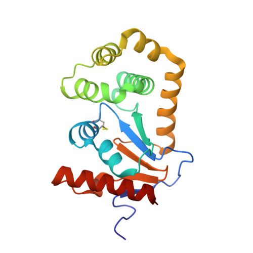
7LHP

Crystal Structure of EcDsbA in a complex with methyl 2-(6-bromo-2-phenylbenzofuran-3-yl)acetate


Experimental Data Snapshot

  • Method: X-RAY DIFFRACTION
  • Resolution: 1.90 Å
  • R-Value Free: 0.225 
  • R-Value Work: 0.179 
  • R-Value Observed: 0.181 

wwPDB Validation   3D Report Full Report


Ligand Structure Quality Assessment 


This is version 1.2 of the entry. See complete history


Literature

Elaboration of a benzofuran scaffold and evaluation of binding affinity and inhibition of Escherichia coli DsbA: A fragment-based drug design approach to novel antivirulence compounds.

Duncan, L.F.Wang, G.Ilyichova, O.V.Dhouib, R.Totsika, M.Scanlon, M.J.Heras, B.Abbott, B.M.

(2021) Bioorg Med Chem 45: 116315-116315

  • DOI: https://doi.org/10.1016/j.bmc.2021.116315
  • Primary Citation of Related Structures:  
    6XSP, 6XSQ, 6XT3, 7L76, 7L7C, 7LHP

  • PubMed Abstract: 

    Bacterial thiol-disulfide oxidoreductase DsbA is essential for bacterial virulence factor assembly and has been identified as a viable antivirulence target. Herein, we report a structure-based elaboration of a benzofuran hit that bound to the active site groove of Escherichia coli DsbA. Substituted phenyl groups were installed at the 5- and 6-position of the benzofuran using Suzuki-Miyaura coupling. HSQC NMR titration experiments showed dissociation constants of this series in the high µM to low mM range and X-ray crystallography produced three co-structures, showing binding in the hydrophobic groove, comparable with that of the previously reported benzofurans. The 6-(m-methoxy)phenyl analogue (2b), which showed a promising binding pose, was chosen for elaboration from the C-2 position. The 2,6-disubstituted analogues bound to the hydrophobic region of the binding groove and the C-2 groups extended into the more polar, previously un-probed, region of the binding groove. Biochemical analysis of the 2,6-disubsituted analogues showed they inhibited DsbA oxidation activity in vitro. The results indicate the potential to develop the elaborated benzofuran series into a novel class of antivirulence compounds.


  • Organizational Affiliation

    Department of Chemistry and Physics, La Trobe Institute for Molecular Science, La Trobe University, Melbourne, VIC 3086, Australia.


Macromolecules
Find similar proteins by:  (by identity cutoff)  |  3D Structure
Entity ID: 1
MoleculeChains Sequence LengthOrganismDetailsImage
Thiol:disulfide interchange protein DsbA
A, B
189Escherichia coli K-12Mutation(s): 0 
Gene Names: dsbAdsfppfAb3860JW3832
UniProt
Find proteins for P0AEG4 (Escherichia coli (strain K12))
Explore P0AEG4 
Go to UniProtKB:  P0AEG4
Entity Groups  
Sequence Clusters30% Identity50% Identity70% Identity90% Identity95% Identity100% Identity
UniProt GroupP0AEG4
Sequence Annotations
Expand
  • Reference Sequence
Small Molecules
Ligands 2 Unique
IDChains Name / Formula / InChI Key2D Diagram3D Interactions
Y1G (Subject of Investigation/LOI)
Query on Y1G

Download Ideal Coordinates CCD File 
C [auth A](6-bromo-2-phenyl-1-benzofuran-3-yl)acetic acid
C16 H11 Br O3
GRIUNLJADADECO-UHFFFAOYSA-N
CU
Query on CU

Download Ideal Coordinates CCD File 
D [auth B]COPPER (II) ION
Cu
JPVYNHNXODAKFH-UHFFFAOYSA-N
Experimental Data & Validation

Experimental Data

  • Method: X-RAY DIFFRACTION
  • Resolution: 1.90 Å
  • R-Value Free: 0.225 
  • R-Value Work: 0.179 
  • R-Value Observed: 0.181 
  • Space Group: C 1 2 1
Unit Cell:
Length ( Å )Angle ( ˚ )
a = 118.211α = 90
b = 63.803β = 125.705
c = 74.767γ = 90
Software Package:
Software NamePurpose
PHENIXrefinement
XDSdata reduction
Aimlessdata scaling
PHASERphasing

Structure Validation

View Full Validation Report


Ligand Structure Quality Assessment 


Entry History 

Deposition Data

Revision History  (Full details and data files)

  • Version 1.0: 2021-08-11
    Type: Initial release
  • Version 1.1: 2021-08-25
    Changes: Database references
  • Version 1.2: 2023-10-18
    Changes: Data collection, Refinement description