7LEP

The composite LBD-TMD structure combined from all hippocampal AMPAR subtypes at 3.25 Angstrom resolution


Experimental Data Snapshot

  • Method: ELECTRON MICROSCOPY
  • Resolution: 3.25 Å
  • Aggregation State: PARTICLE 
  • Reconstruction Method: SINGLE PARTICLE 

wwPDB Validation   3D Report Full Report


This is version 1.2 of the entry. See complete history


Literature

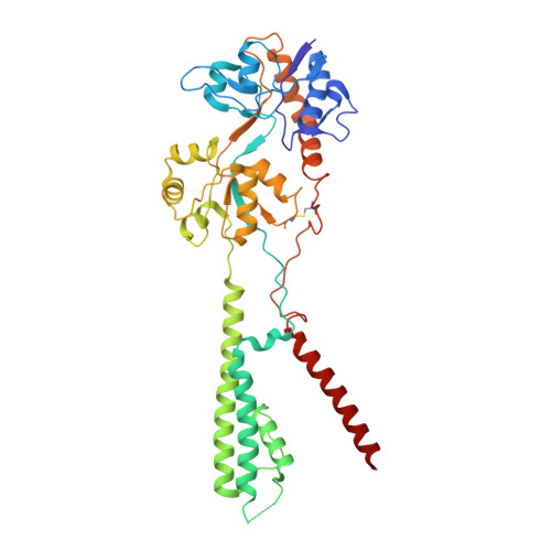
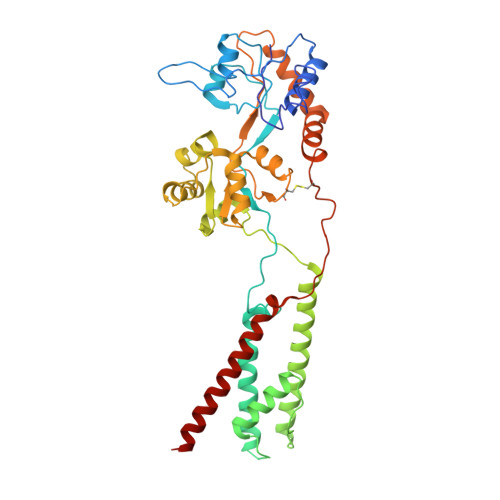
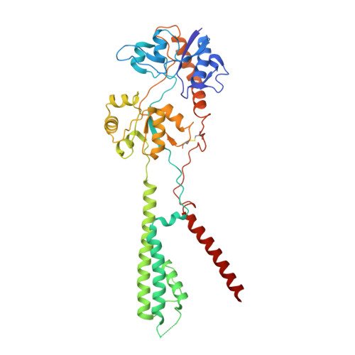
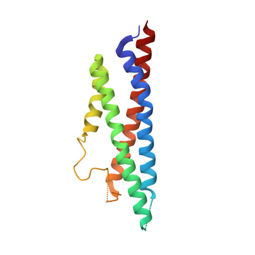
Hippocampal AMPA receptor assemblies and mechanism of allosteric inhibition.

Yu, J.Rao, P.Clark, S.Mitra, J.Ha, T.Gouaux, E.

(2021) Nature 594: 448-453

  • DOI: https://doi.org/10.1038/s41586-021-03540-0
  • Primary Citation of Related Structures:  
    7LDD, 7LDE, 7LEP

  • PubMed Abstract: 

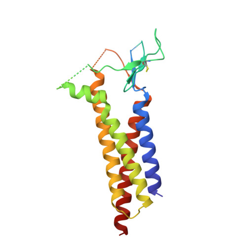
    AMPA-selective glutamate receptors mediate the transduction of signals between the neuronal circuits of the hippocampus 1 . The trafficking, localization, kinetics and pharmacology of AMPA receptors are tuned by an ensemble of auxiliary protein subunits, which are integral membrane proteins that associate with the receptor to yield bona fide receptor signalling complexes 2 . Thus far, extensive studies of recombinant AMPA receptor-auxiliary subunit complexes using engineered protein constructs have not been able to faithfully elucidate the molecular architecture of hippocampal AMPA receptor complexes. Here we obtain mouse hippocampal, calcium-impermeable AMPA receptor complexes using immunoaffinity purification and use single-molecule fluorescence and cryo-electron microscopy experiments to elucidate three major AMPA receptor-auxiliary subunit complexes. The GluA1-GluA2, GluA1-GluA2-GluA3 and GluA2-GluA3 receptors are the predominant assemblies, with the auxiliary subunits TARP-γ8 and CNIH2-SynDIG4 non-stochastically positioned at the B'/D' and A'/C' positions, respectively. We further demonstrate how the receptor-TARP-γ8 stoichiometry explains the mechanism of and submaximal inhibition by a clinically relevant, brain-region-specific allosteric inhibitor.


  • Organizational Affiliation

    Vollum Institute, Oregon Health & Science University, Portland, OR, USA.


Macromolecules
Find similar proteins by:  (by identity cutoff)  |  3D Structure
Entity ID: 1
MoleculeChains Sequence LengthOrganismDetailsImage
Mix of AMPAR subunits (GluA1, GluA3, and GluA4)414Mus musculusMutation(s): 0 
Entity Groups  
Sequence Clusters30% Identity50% Identity70% Identity90% Identity95% Identity100% Identity
Sequence Annotations
Expand
  • Reference Sequence
Find similar proteins by:  (by identity cutoff)  |  3D Structure
Entity ID: 2
MoleculeChains Sequence LengthOrganismDetailsImage
Glutamate receptor 2
B, D
424Mus musculusMutation(s): 0 
Membrane Entity: Yes 
UniProt & NIH Common Fund Data Resources
Find proteins for P23819 (Mus musculus)
Explore P23819 
Go to UniProtKB:  P23819
IMPC:  MGI:95809
Entity Groups  
Sequence Clusters30% Identity50% Identity70% Identity90% Identity95% Identity100% Identity
UniProt GroupP23819
Sequence Annotations
Expand
  • Reference Sequence
Find similar proteins by:  (by identity cutoff)  |  3D Structure
Entity ID: 3
MoleculeChains Sequence LengthOrganismDetailsImage
Mix of AMPAR subunits (GluA1, GluA3, and GluAX)413Mus musculusMutation(s): 0 
Entity Groups  
Sequence Clusters30% Identity50% Identity70% Identity90% Identity95% Identity100% Identity
Sequence Annotations
Expand
  • Reference Sequence
Find similar proteins by:  (by identity cutoff)  |  3D Structure
Entity ID: 4
MoleculeChains Sequence LengthOrganismDetailsImage
Protein cornichon homolog 2
E, F
159Mus musculusMutation(s): 0 
Membrane Entity: Yes 
UniProt & NIH Common Fund Data Resources
Find proteins for O35089 (Mus musculus)
Explore O35089 
Go to UniProtKB:  O35089
IMPC:  MGI:1277225
Entity Groups  
Sequence Clusters30% Identity50% Identity70% Identity90% Identity95% Identity100% Identity
UniProt GroupO35089
Sequence Annotations
Expand
  • Reference Sequence
Find similar proteins by:  (by identity cutoff)  |  3D Structure
Entity ID: 5
MoleculeChains Sequence LengthOrganismDetailsImage
Voltage-dependent calcium channel gamma-8 subunit
G, H
215Mus musculusMutation(s): 0 
Membrane Entity: Yes 
UniProt
Find proteins for Q8VHW2 (Mus musculus)
Explore Q8VHW2 
Go to UniProtKB:  Q8VHW2
Entity Groups  
Sequence Clusters30% Identity50% Identity70% Identity90% Identity95% Identity100% Identity
UniProt GroupQ8VHW2
Sequence Annotations
Expand
  • Reference Sequence
Small Molecules
Ligands 7 Unique
IDChains Name / Formula / InChI Key2D Diagram3D Interactions
POV
Query on POV

Download Ideal Coordinates CCD File 
AA [auth C]
BA [auth C]
CA [auth C]
DA [auth C]
EA [auth C]
AA [auth C],
BA [auth C],
CA [auth C],
DA [auth C],
EA [auth C],
FA [auth C],
GA [auth C],
IA [auth D],
J [auth A],
JA [auth D],
K [auth A],
KA [auth D],
L [auth A],
LA [auth D],
M [auth A],
MA [auth D],
N [auth A],
NA [auth D],
O [auth A],
OA [auth E],
P [auth A],
PA [auth F],
Q [auth A],
S [auth B],
TA [auth G],
U [auth B],
V [auth B],
W [auth B],
X [auth B],
XA [auth H]
(2S)-3-(hexadecanoyloxy)-2-[(9Z)-octadec-9-enoyloxy]propyl 2-(trimethylammonio)ethyl phosphate
C42 H82 N O8 P
WTJKGGKOPKCXLL-PFDVCBLKSA-N
ZK1
Query on ZK1

Download Ideal Coordinates CCD File 
HA [auth D],
I [auth A],
R [auth B],
Y [auth C]
{[7-morpholin-4-yl-2,3-dioxo-6-(trifluoromethyl)-3,4-dihydroquinoxalin-1(2H)-yl]methyl}phosphonic acid
C14 H15 F3 N3 O6 P
WZMQMKNCWDCCMT-UHFFFAOYSA-N
XVD (Subject of Investigation/LOI)
Query on XVD

Download Ideal Coordinates CCD File 
QA [auth G],
UA [auth H]
6-[2-chloro-6-(trifluoromethoxy)phenyl]-1H-benzimidazol-2-ol
C14 H8 Cl F3 N2 O2
COBXSLRIXGQVGS-UHFFFAOYSA-N
C14
Query on C14

Download Ideal Coordinates CCD File 
T [auth B]TETRADECANE
C14 H30
BGHCVCJVXZWKCC-UHFFFAOYSA-N
D12
Query on D12

Download Ideal Coordinates CCD File 
SA [auth G],
WA [auth H]
DODECANE
C12 H26
SNRUBQQJIBEYMU-UHFFFAOYSA-N
D10
Query on D10

Download Ideal Coordinates CCD File 
Z [auth C]DECANE
C10 H22
DIOQZVSQGTUSAI-UHFFFAOYSA-N
OCT
Query on OCT

Download Ideal Coordinates CCD File 
RA [auth G],
VA [auth H]
N-OCTANE
C8 H18
TVMXDCGIABBOFY-UHFFFAOYSA-N
Experimental Data & Validation

Experimental Data

  • Method: ELECTRON MICROSCOPY
  • Resolution: 3.25 Å
  • Aggregation State: PARTICLE 
  • Reconstruction Method: SINGLE PARTICLE 

Structure Validation

View Full Validation Report


Entry History & Funding Information

Deposition Data


Funding OrganizationLocationGrant Number
National Institutes of Health/National Institute of Neurological Disorders and Stroke (NIH/NINDS)United StatesR01NS038631

Revision History  (Full details and data files)

  • Version 1.0: 2021-05-12
    Type: Initial release
  • Version 1.1: 2021-05-26
    Changes: Database references
  • Version 1.2: 2021-06-30
    Changes: Database references