7LCV | pdb_00007lcv

Factor H enhancing human antibody fragment (Fab) to meningococcal Factor H binding protein


Experimental Data Snapshot

  • Method: X-RAY DIFFRACTION
  • Resolution: 1.70 Å
  • R-Value Free: 
    0.270 (Depositor), 0.271 (DCC) 
  • R-Value Work: 
    0.239 (Depositor), 0.242 (DCC) 
  • R-Value Observed: 
    0.240 (Depositor) 

Starting Model: experimental
View more details

wwPDB Validation 3D Report Full Report

Validation slider image for 7LCV

This is version 1.3 of the entry. See complete history

Literature

Two human antibodies to a meningococcal serogroup B vaccine antigen enhance binding of complement Factor H by stabilizing the Factor H binding site.

Sands, N.A.Beernink, P.T.

(2021) PLoS Pathog 17: e1009655-e1009655

  • DOI: https://doi.org/10.1371/journal.ppat.1009655
  • Primary Citation Related Structures: 
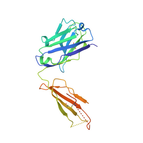
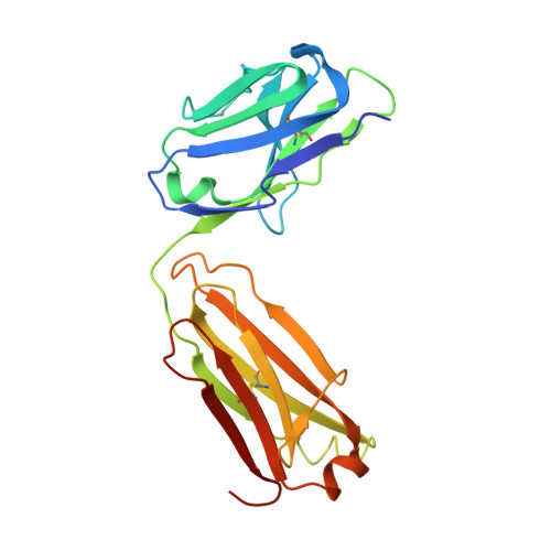
    7KE1, 7KET, 7LCV

  • PubMed Abstract: 

    Microbial pathogens bind host complement regulatory proteins to evade the immune system. The bacterial pathogen Neisseria meningitidis, or meningococcus, binds several complement regulators, including human Factor H (FH). FH binding protein (FHbp) is a component of two licensed meningococcal vaccines and in mice FHbp elicits antibodies that inhibit binding of FH to FHbp, which defeat the bacterial evasion mechanism. However, humans vaccinated with FHbp develop antibodies that enhance binding of FH to the bacteria, which could limit the effectiveness of the vaccines. In the present study, we show that two vaccine-elicited antibody fragments (Fabs) isolated from different human subjects increase binding of complement FH to meningococcal FHbp by ELISA. The two Fabs have different effects on the kinetics of FH binding to immobilized FHbp as measured by surface plasmon resonance. The 1.7- and 2.0-Å resolution X-ray crystal structures of the Fabs in complexes with FHbp illustrate that the two Fabs bind to similar epitopes on the amino-terminal domain of FHbp, adjacent to the FH binding site. Superposition models of ternary complexes of each Fab with FHbp and FH show that there is likely minimal contact between the Fabs and FH. Collectively, the structures reveal that the Fabs enhance binding of FH to FHbp by altering the conformations and mobilities of two loops adjacent to the FH binding site of FHbp. In addition, the 1.5 Å-resolution structure of one of the isolated Fabs defines the structural rearrangements associated with binding to FHbp. The FH-enhancing human Fabs, which are mirrored in the human polyclonal antibody responses, have important implications for tuning the effectiveness of FHbp-based vaccines.


  • Organizational Affiliation
    • Division of Infectious Diseases and Global Health, Department of Pediatrics, School of Medicine, University of California San Francisco, San Francisco, California, United States of America.

Macromolecule Content 

  • Total Structure Weight: 75.69 kDa 
  • Atom Count: 5,032 
  • Modeled Residue Count: 628 
  • Deposited Residue Count: 707 
  • Unique protein chains: 3

Macromolecules

Find similar proteins by:|  3D Structure
Entity ID: 1
MoleculeChains  Sequence LengthOrganismDetailsImage
Immunoglobulin heavy chain Fd fragment227Homo sapiensMutation(s): 0 
Find similar proteins by:|  3D Structure
Entity ID: 2
MoleculeChains  Sequence LengthOrganismDetailsImage
Immunoglobulin kappa light chain217Homo sapiensMutation(s): 0 
Find similar proteins by:|  3D Structure
Entity ID: 3
MoleculeChains  Sequence LengthOrganismDetailsImage
Factor H binding protein263Neisseria meningitidisMutation(s): 0 
Gene Names: fhbp
UniProt
Find proteins for Q9JXV4 (Neisseria meningitidis serogroup B (strain ATCC BAA-335 / MC58))
Explore Q9JXV4 
Go to UniProtKB:  Q9JXV4
Entity Groups
Sequence Clusters30% Identity50% Identity70% Identity90% Identity95% Identity100% Identity
UniProt GroupQ9JXV4
Sequence Annotations
Expand
Reference Sequence

Experimental Data & Validation

Experimental Data

  • Method: X-RAY DIFFRACTION
  • Resolution: 1.70 Å
  • R-Value Free:  0.270 (Depositor), 0.271 (DCC) 
  • R-Value Work:  0.239 (Depositor), 0.242 (DCC) 
  • R-Value Observed: 0.240 (Depositor) 
Space Group: P 1
Unit Cell:
Length ( Å )Angle ( ˚ )
a = 39.47α = 73.781
b = 52.37β = 84.482
c = 84.56γ = 74.173
Software Package:
Software NamePurpose
PHENIXrefinement
XDSdata reduction
XSCALEdata scaling
PHASERphasing

Structure Validation

View Full Validation Report


Entry History 

& Funding Information

Deposition Data


Funding OrganizationLocationGrant Number
National Institutes of Health/National Institute Of Allergy and Infectious Diseases (NIH/NIAID)United States1R01AI134868

Revision History  (Full details and data files)

  • Version 1.0: 2021-06-09
    Type: Initial release
  • Version 1.1: 2021-07-14
    Changes: Database references
  • Version 1.2: 2023-10-18
    Changes: Data collection, Database references, Refinement description
  • Version 1.3: 2024-11-20
    Changes: Structure summary