


Crystal structure of the tandem bromodomain (BD1, BD2) of human TAF1 bound to ZS1-585
Karim, M.R., Schonbrunn, E.To be published.
Experimental Data Snapshot
Starting Model: experimental
View more details
Entity ID: 1 | |||||
|---|---|---|---|---|---|
| Molecule | Chains | Sequence Length | Organism | Details | Image |
| Transcription initiation factor TFIID subunit 1 | 265 | Homo sapiens | Mutation(s): 0 Gene Names: TAF1, BA2R, CCG1, CCGS, TAF2A EC: 2.3.1.48 (PDB Primary Data), 2.7.11.1 (PDB Primary Data) | 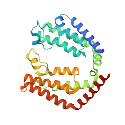 | |
UniProt & NIH Common Fund Data Resources | |||||
PHAROS: P21675 GTEx: ENSG00000147133 | |||||
Entity Groups | |||||
| Sequence Clusters | 30% Identity50% Identity70% Identity90% Identity95% Identity100% Identity | ||||
| UniProt Group | P21675 | ||||
Sequence AnnotationsExpand | |||||
Reference Sequence | |||||
| Ligands 3 Unique | |||||
|---|---|---|---|---|---|
| ID | Chains | Name / Formula / InChI Key | 2D Diagram | 3D Interactions | |
| XWV (Subject of Investigation/LOI) Download:Ideal Coordinates CCD File | U [auth A] | 5-{4-[(3R)-3-methylmorpholin-4-yl]-6-[1-(S-methylsulfonimidoyl)cyclopropyl]pyrimidin-2-yl}isoquinoline C22 H25 N5 O2 S QKKJIBZPNHULHG-DHMKHTPVSA-N |  | ||
| PEG Download:Ideal Coordinates CCD File | K [auth A] | DI(HYDROXYETHYL)ETHER C4 H10 O3 MTHSVFCYNBDYFN-UHFFFAOYSA-N |  | ||
| EDO Download:Ideal Coordinates CCD File | B [auth A] C [auth A] D [auth A] E [auth A] F [auth A] | 1,2-ETHANEDIOL C2 H6 O2 LYCAIKOWRPUZTN-UHFFFAOYSA-N |  | ||
| Length ( Å ) | Angle ( ˚ ) |
|---|---|
| a = 45.167 | α = 90 |
| b = 54.665 | β = 90 |
| c = 122.049 | γ = 90 |
| Software Name | Purpose |
|---|---|
| PHENIX | refinement |
| XDS | data reduction |
| Aimless | data scaling |
| PHASER | phasing |


