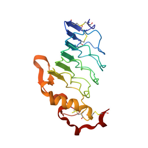
7LA8

O6 variable lymphocyte receptor ectodomain bound to 3-HSO3-Gal-4GlcNAc


Experimental Data Snapshot

  • Method: X-RAY DIFFRACTION
  • Resolution: 1.90 Å
  • R-Value Free: 0.229 
  • R-Value Work: 0.184 
  • R-Value Observed: 0.186 

wwPDB Validation   3D Report Full Report


Ligand Structure Quality Assessment 


This is version 1.1 of the entry. See complete history


Literature

Novel lamprey antibody recognizes terminal sulfated galactose epitopes on mammalian glycoproteins.

McKitrick, T.R.Bernard, S.M.Noll, A.J.Collins, B.C.Goth, C.K.McQuillan, A.M.Heimburg-Molinaro, J.Herrin, B.R.Wilson, I.A.Cooper, M.D.Cummings, R.D.

(2021) Commun Biol 4: 674-674

  • DOI: https://doi.org/10.1038/s42003-021-02199-7
  • Primary Citation of Related Structures:  
    7LA7, 7LA8

  • PubMed Abstract: 

    The terminal galactose residues of N- and O-glycans in animal glycoproteins are often sialylated and/or fucosylated, but sulfation, such as 3-O-sulfated galactose (3-O-SGal), represents an additional, but poorly understood modification. To this end, we have developed a novel sea lamprey variable lymphocyte receptor (VLR) termed O6 to explore 3-O-SGal expression. O6 was engineered as a recombinant murine IgG chimera and its specificity and affinity to the 3-O-SGal epitope was defined using a variety of approaches, including glycan and glycoprotein microarray analyses, isothermal calorimetry, ligand-bound crystal structure, FACS, and immunohistochemistry of human tissue macroarrays. 3-O-SGal is expressed on N-glycans of many plasma and tissue glycoproteins, but recognition by O6 is often masked by sialic acid and thus exposed by treatment with neuraminidase. O6 recognizes many human tissues, consistent with expression of the cognate sulfotransferases (GAL3ST-2 and GAL3ST-3). The availability of O6 for exploring 3-O-SGal expression could lead to new biomarkers for disease and aid in understanding the functional roles of terminal modifications of glycans and relationships between terminal sulfation, sialylation and fucosylation.


  • Organizational Affiliation

    Department of Surgery, Beth Israel Deaconess Medical Center, Harvard Medical School, Boston, MA, USA.


Macromolecules
Find similar proteins by:  (by identity cutoff)  |  3D Structure
Entity ID: 1
MoleculeChains Sequence LengthOrganismDetailsImage
O6 variable lymphocyte receptor
A, B
173Petromyzon marinusMutation(s): 0 
Entity Groups  
Sequence Clusters30% Identity50% Identity70% Identity90% Identity95% Identity100% Identity
Sequence Annotations
Expand
  • Reference Sequence
Oligosaccharides

Help

Entity ID: 2
MoleculeChains Length2D Diagram Glycosylation3D Interactions
3-O-sulfo-beta-D-galactopyranose-(1-4)-2-acetamido-2-deoxy-beta-D-glucopyranose
C, D
2N/A
Glycosylation Resources
GlyTouCan:  G62117RR
GlyCosmos:  G62117RR
GlyGen:  G62117RR
Experimental Data & Validation

Experimental Data

  • Method: X-RAY DIFFRACTION
  • Resolution: 1.90 Å
  • R-Value Free: 0.229 
  • R-Value Work: 0.184 
  • R-Value Observed: 0.186 
  • Space Group: I 4
Unit Cell:
Length ( Å )Angle ( ˚ )
a = 107.255α = 90
b = 107.255β = 90
c = 63.197γ = 90
Software Package:
Software NamePurpose
PHENIXrefinement
XDSdata reduction
Aimlessdata scaling
PHASERphasing

Structure Validation

View Full Validation Report


Ligand Structure Quality Assessment 


Entry History & Funding Information

Deposition Data


Funding OrganizationLocationGrant Number
National Institutes of Health/National Institute Of Allergy and Infectious Diseases (NIH/NIAID)United States--

Revision History  (Full details and data files)

  • Version 1.0: 2021-07-14
    Type: Initial release
  • Version 1.1: 2023-10-18
    Changes: Data collection, Database references, Refinement description