


Structure of human GGT1 in complex with Lnt1-172 compound.
Terzyan, S.S., Hanigan, M.To be published.
Experimental Data Snapshot
Starting Model: experimental
View more details
Entity ID: 1 | |||||
|---|---|---|---|---|---|
| Molecule | Chains | Sequence Length | Organism | Details | Image |
| Glutathione hydrolase 1 heavy chain | 353 | Homo sapiens | Mutation(s): 1 Gene Names: GGT1, GGT EC: 3.4.19.13 (PDB Primary Data), 2.3.2.2 (PDB Primary Data), 3.4.19.14 (PDB Primary Data) | 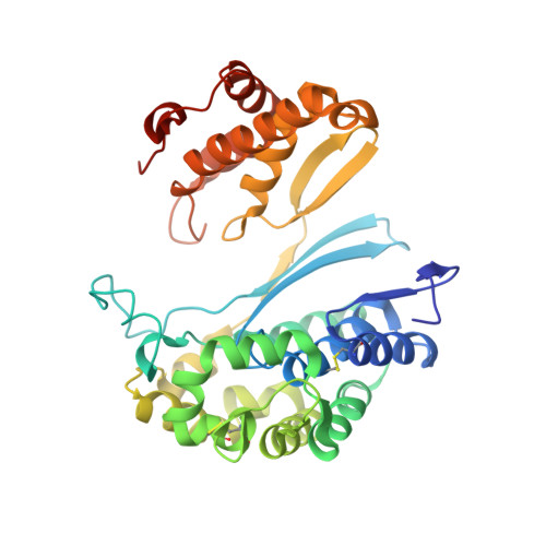 | |
UniProt & NIH Common Fund Data Resources | |||||
PHAROS: P19440 GTEx: ENSG00000100031 | |||||
Entity Groups | |||||
| Sequence Clusters | 30% Identity50% Identity70% Identity90% Identity95% Identity100% Identity | ||||
| UniProt Group | P19440 | ||||
Glycosylation | |||||
| Glycosylation Sites: 4 | Go to GlyGen: P19440-1 | ||||
Sequence AnnotationsExpand | |||||
Reference Sequence | |||||
Entity ID: 2 | |||||
|---|---|---|---|---|---|
| Molecule | Chains | Sequence Length | Organism | Details | Image |
| Glutathione hydrolase 1 light chain | 189 | Homo sapiens | Mutation(s): 0 Gene Names: GGT1, GGT EC: 3.4.19.13 (PDB Primary Data), 2.3.2.2 (PDB Primary Data), 3.4.19.14 (PDB Primary Data) | 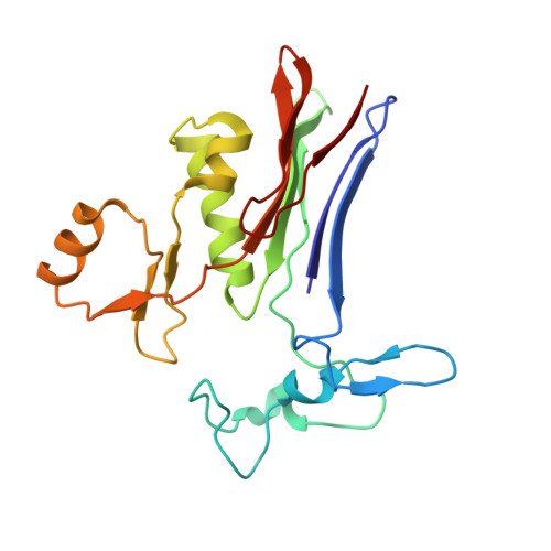 | |
UniProt & NIH Common Fund Data Resources | |||||
PHAROS: P19440 GTEx: ENSG00000100031 | |||||
Entity Groups | |||||
| Sequence Clusters | 30% Identity50% Identity70% Identity90% Identity95% Identity100% Identity | ||||
| UniProt Group | P19440 | ||||
Glycosylation | |||||
| Glycosylation Sites: 1 | Go to GlyGen: P19440-1 | ||||
Sequence AnnotationsExpand | |||||
Reference Sequence | |||||
| Ligands 4 Unique | |||||
|---|---|---|---|---|---|
| ID | Chains | Name / Formula / InChI Key | 2D Diagram | 3D Interactions | |
| EPE Download:Ideal Coordinates CCD File | G [auth A] | 4-(2-HYDROXYETHYL)-1-PIPERAZINE ETHANESULFONIC ACID C8 H18 N2 O4 S JKMHFZQWWAIEOD-UHFFFAOYSA-N |  | ||
| XSM (Subject of Investigation/LOI) Download:Ideal Coordinates CCD File | J [auth B] | (2R)-4-borono-2-{[(1H-imidazol-4-yl)methyl]amino}butanoic acid C8 H14 B N3 O4 JBISBMUZBNLAPG-SSDOTTSWSA-N |  | ||
| NAG Download:Ideal Coordinates CCD File | C [auth A], D [auth A], E [auth A], F [auth A], I [auth B] | 2-acetamido-2-deoxy-beta-D-glucopyranose C8 H15 N O6 OVRNDRQMDRJTHS-FMDGEEDCSA-N |  | ||
| CL Download:Ideal Coordinates CCD File | H [auth A] | CHLORIDE ION Cl VEXZGXHMUGYJMC-UHFFFAOYSA-M |  | ||
| Length ( Å ) | Angle ( ˚ ) |
|---|---|
| a = 103.223 | α = 90 |
| b = 118.855 | β = 90 |
| c = 101.457 | γ = 90 |
| Software Name | Purpose |
|---|---|
| REFMAC | refinement |
| PDB_EXTRACT | data extraction |
| HKL-2000 | data reduction |
| HKL-2000 | data scaling |
| REFMAC | phasing |
| Funding Organization | Location | Grant Number |
|---|---|---|
| National Institutes of Health/National Institute of General Medical Sciences (NIH/NIGMS) | United States | R01GM125952 |


