7L5I

Crystal Structure of Haemophilus influenzae MtsZ at pH 7.0


Experimental Data Snapshot

  • Method: X-RAY DIFFRACTION
  • Resolution: 1.73 Å
  • R-Value Free: 0.160 
  • R-Value Work: 0.132 
  • R-Value Observed: 0.133 

wwPDB Validation   3D Report Full Report


Ligand Structure Quality Assessment 


This is version 1.3 of the entry. See complete history


Literature

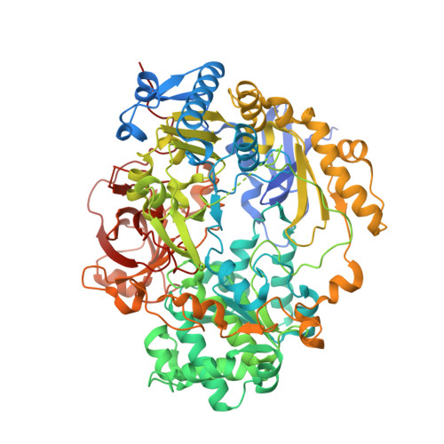
Active site architecture reveals coordination sphere flexibility and specificity determinants in a group of closely related molybdoenzymes.

Struwe, M.A.Kalimuthu, P.Luo, Z.Zhong, Q.Ellis, D.Yang, J.Khadanand, K.C.Harmer, J.R.Kirk, M.L.McEwan, A.G.Clement, B.Bernhardt, P.V.Kobe, B.Kappler, U.

(2021) J Biol Chem 296: 100672-100672

  • DOI: https://doi.org/10.1016/j.jbc.2021.100672
  • Primary Citation of Related Structures:  
    7L5I, 7L5S

  • PubMed Abstract: 

    MtsZ is a molybdenum-containing methionine sulfoxide reductase that supports virulence in the human respiratory pathogen Haemophilus influenzae (Hi). HiMtsZ belongs to a group of structurally and spectroscopically uncharacterized S-/N-oxide reductases, all of which are found in bacterial pathogens. Here, we have solved the crystal structure of HiMtsZ, which reveals that the HiMtsZ substrate-binding site encompasses a previously unrecognized part that accommodates the methionine sulfoxide side chain via interaction with His182 and Arg166. Charge and amino acid composition of this side chain-binding region vary and, as indicated by electrochemical, kinetic, and docking studies, could explain the diverse substrate specificity seen in closely related enzymes of this type. The HiMtsZ Mo active site has an underlying structural flexibility, where dissociation of the central Ser187 ligand affected catalysis at low pH. Unexpectedly, the two main HiMtsZ electron paramagnetic resonance (EPR) species resembled not only a related dimethyl sulfoxide reductase but also a structurally unrelated nitrate reductase that possesses an Asp-Mo ligand. This suggests that contrary to current views, the geometry of the Mo center and its primary ligands, rather than the specific amino acid environment, is the main determinant of the EPR properties of mononuclear Mo enzymes. The flexibility in the electronic structure of the Mo centers is also apparent in two of three HiMtsZ EPR-active Mo(V) species being catalytically incompetent off-pathway forms that could not be fully oxidized.


  • Organizational Affiliation

    Australian Infectious Disease Research Centre, School of Chemistry and Molecular Biosciences, The University of Queensland, Brisbane, Qld, Australia; Pharmazeutisches Institut, Christian-Albrechts-Universität Kiel, Kiel, Germany.


Macromolecules
Find similar proteins by:  (by identity cutoff)  |  3D Structure
Entity ID: 1
MoleculeChains Sequence LengthOrganismDetailsImage
Trimethylamine-N-oxide reductase813Haemophilus influenzaeMutation(s): 0 
Gene Names: torZBV064_00672
EC: 1.7.2.3
UniProt
Find proteins for P44798 (Haemophilus influenzae (strain ATCC 51907 / DSM 11121 / KW20 / Rd))
Explore P44798 
Go to UniProtKB:  P44798
Entity Groups  
Sequence Clusters30% Identity50% Identity70% Identity90% Identity95% Identity100% Identity
UniProt GroupP44798
Sequence Annotations
Expand
  • Reference Sequence
Small Molecules
Ligands 5 Unique
IDChains Name / Formula / InChI Key2D Diagram3D Interactions
MGD (Subject of Investigation/LOI)
Query on MGD

Download Ideal Coordinates CCD File 
D [auth A],
E [auth A]
2-AMINO-5,6-DIMERCAPTO-7-METHYL-3,7,8A,9-TETRAHYDRO-8-OXA-1,3,9,10-TETRAAZA-ANTHRACEN-4-ONE GUANOSINE DINUCLEOTIDE
C20 H26 N10 O13 P2 S2
VQAGYJCYOLHZDH-ILXWUORBSA-N
EPE
Query on EPE

Download Ideal Coordinates CCD File 
B [auth A],
C [auth A],
H [auth A]
4-(2-HYDROXYETHYL)-1-PIPERAZINE ETHANESULFONIC ACID
C8 H18 N2 O4 S
JKMHFZQWWAIEOD-UHFFFAOYSA-N
MO (Subject of Investigation/LOI)
Query on MO

Download Ideal Coordinates CCD File 
F [auth A]MOLYBDENUM ATOM
Mo
ZOKXTWBITQBERF-UHFFFAOYSA-N
CL
Query on CL

Download Ideal Coordinates CCD File 
G [auth A]CHLORIDE ION
Cl
VEXZGXHMUGYJMC-UHFFFAOYSA-M
O (Subject of Investigation/LOI)
Query on O

Download Ideal Coordinates CCD File 
I [auth A]OXYGEN ATOM
O
XLYOFNOQVPJJNP-UHFFFAOYSA-N
Experimental Data & Validation

Experimental Data

  • Method: X-RAY DIFFRACTION
  • Resolution: 1.73 Å
  • R-Value Free: 0.160 
  • R-Value Work: 0.132 
  • R-Value Observed: 0.133 
  • Space Group: P 21 21 21
Unit Cell:
Length ( Å )Angle ( ˚ )
a = 58.2α = 90
b = 93.681β = 90
c = 132.777γ = 90
Software Package:
Software NamePurpose
PHENIXrefinement
XDSdata reduction
Aimlessdata scaling
MOLREPphasing
RESOLVEmodel building
PDB_EXTRACTdata extraction

Structure Validation

View Full Validation Report


Ligand Structure Quality Assessment 


Entry History & Funding Information

Deposition Data


Funding OrganizationLocationGrant Number
National Health and Medical Research Council (NHMRC, Australia)AustraliaGNT1158451
National Health and Medical Research Council (NHMRC, Australia)Australia1180826

Revision History  (Full details and data files)

  • Version 1.0: 2021-04-21
    Type: Initial release
  • Version 1.1: 2021-05-05
    Changes: Database references
  • Version 1.2: 2021-07-21
    Changes: Database references
  • Version 1.3: 2023-10-18
    Changes: Data collection, Database references, Refinement description