7KO5

Structure of cardiac native thin filament at pCa=5.8 having upper and lower troponins in Ca2+ bound state

  • Classification: CONTRACTILE PROTEIN
  • Organism(s): Sus scrofa
  • Mutation(s): No 

  • Deposited: 2020-11-06 Released: 2021-03-24 
  • Deposition Author(s): Galkin, V.E., Risi, C.M.
  • Funding Organization(s): National Institutes of Health/National Heart, Lung, and Blood Institute (NIH/NHLBI), National Institutes of Health/National Institute of General Medical Sciences (NIH/NIGMS)

Experimental Data Snapshot

  • Method: ELECTRON MICROSCOPY
  • Resolution: 7.80 Å
  • Aggregation State: FILAMENT 
  • Reconstruction Method: SINGLE PARTICLE 

wwPDB Validation   3D Report Full Report


This is version 1.2 of the entry. See complete history


Literature

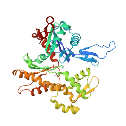
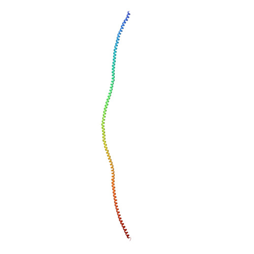
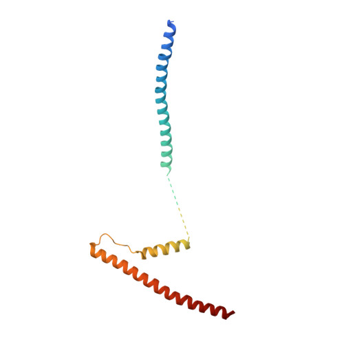
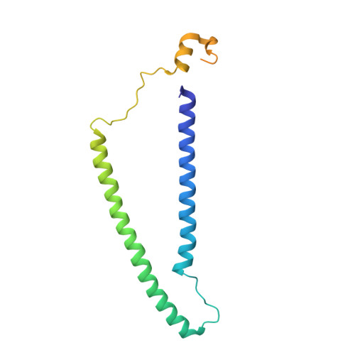
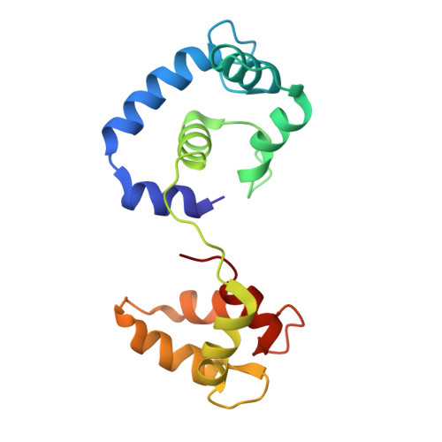
The structure of the native cardiac thin filament at systolic Ca 2+ levels.

Risi, C.M.Pepper, I.Belknap, B.Landim-Vieira, M.White, H.D.Dryden, K.Pinto, J.R.Chase, P.B.Galkin, V.E.

(2021) Proc Natl Acad Sci U S A 118

  • DOI: https://doi.org/10.1073/pnas.2024288118
  • Primary Citation of Related Structures:  
    7KO4, 7KO5, 7KO7, 7KON, 7KOR

  • PubMed Abstract: 

    Every heartbeat relies on cyclical interactions between myosin thick and actin thin filaments orchestrated by rising and falling Ca 2+ levels. Thin filaments are comprised of two actin strands, each harboring equally separated troponin complexes, which bind Ca 2+ to move tropomyosin cables away from the myosin binding sites and, thus, activate systolic contraction. Recently, structures of thin filaments obtained at low (pCa ∼9) or high (pCa ∼3) Ca 2+ levels revealed the transition between the Ca 2+ -free and Ca 2+ -bound states. However, in working cardiac muscle, Ca 2+ levels fluctuate at intermediate values between pCa ∼6 and pCa ∼7. The structure of the thin filament at physiological Ca 2+ levels is unknown. We used cryoelectron microscopy and statistical analysis to reveal the structure of the cardiac thin filament at systolic pCa = 5.8. We show that the two strands of the thin filament consist of a mixture of regulatory units, which are composed of Ca 2+ -free, Ca 2+ -bound, or mixed (e.g., Ca 2+ free on one side and Ca 2+ bound on the other side) troponin complexes. We traced troponin complex conformations along and across individual thin filaments to directly determine the structural composition of the cardiac native thin filament at systolic Ca 2+ levels. We demonstrate that the two thin filament strands are activated stochastically with short-range cooperativity evident only on one of the two strands. Our findings suggest a mechanism by which cardiac muscle is regulated by narrow range Ca 2+ fluctuations.


  • Organizational Affiliation

    Department of Physiological Sciences, Eastern Virginia Medical School, Norfolk, VA 23507.


Macromolecules
Find similar proteins by:  (by identity cutoff)  |  3D Structure
Entity ID: 1
MoleculeChains Sequence LengthOrganismDetailsImage
Actin, alpha skeletal muscle
A, B, C, D, E
A, B, C, D, E, F, G, H, I, J, K, L, M, N, O
375Sus scrofaMutation(s): 0 
UniProt
Find proteins for P68137 (Sus scrofa)
Explore P68137 
Go to UniProtKB:  P68137
Entity Groups  
Sequence Clusters30% Identity50% Identity70% Identity90% Identity95% Identity100% Identity
UniProt GroupP68137
Sequence Annotations
Expand
  • Reference Sequence
Find similar proteins by:  (by identity cutoff)  |  3D Structure
Entity ID: 2
MoleculeChains Sequence LengthOrganismDetailsImage
Tropomyosin alpha-1 chain
P, Q, R, S, W
P, Q, R, S, W, X, Y, Z
286Sus scrofaMutation(s): 0 
UniProt
Find proteins for P42639 (Sus scrofa)
Explore P42639 
Go to UniProtKB:  P42639
Entity Groups  
Sequence Clusters30% Identity50% Identity70% Identity90% Identity95% Identity100% Identity
UniProt GroupP42639
Sequence Annotations
Expand
  • Reference Sequence
Find similar proteins by:  (by identity cutoff)  |  3D Structure
Entity ID: 3
MoleculeChains Sequence LengthOrganismDetailsImage
Troponin T, cardiac muscleAA [auth a],
T
186Sus scrofaMutation(s): 0 
UniProt
Find proteins for A0A4X1SIP4 (Sus scrofa)
Explore A0A4X1SIP4 
Go to UniProtKB:  A0A4X1SIP4
Entity Groups  
Sequence Clusters30% Identity50% Identity70% Identity90% Identity95% Identity100% Identity
UniProt GroupA0A4X1SIP4
Sequence Annotations
Expand
  • Reference Sequence
Find similar proteins by:  (by identity cutoff)  |  3D Structure
Entity ID: 4
MoleculeChains Sequence LengthOrganismDetailsImage
Troponin I, cardiac muscleBA [auth b],
U
170Sus scrofaMutation(s): 0 
Entity Groups  
Sequence Clusters30% Identity50% Identity70% Identity90% Identity95% Identity100% Identity
Sequence Annotations
Expand
  • Reference Sequence
Find similar proteins by:  (by identity cutoff)  |  3D Structure
Entity ID: 5
MoleculeChains Sequence LengthOrganismDetailsImage
Troponin CCA [auth c],
V
160Sus scrofaMutation(s): 0 
UniProt
Find proteins for P63317 (Sus scrofa)
Explore P63317 
Go to UniProtKB:  P63317
Entity Groups  
Sequence Clusters30% Identity50% Identity70% Identity90% Identity95% Identity100% Identity
UniProt GroupP63317
Sequence Annotations
Expand
  • Reference Sequence
Experimental Data & Validation

Experimental Data

  • Method: ELECTRON MICROSCOPY
  • Resolution: 7.80 Å
  • Aggregation State: FILAMENT 
  • Reconstruction Method: SINGLE PARTICLE 
EM Software:
TaskSoftware PackageVersion
MODEL REFINEMENTPHENIX

Structure Validation

View Full Validation Report


Entry History & Funding Information

Deposition Data


Funding OrganizationLocationGrant Number
National Institutes of Health/National Heart, Lung, and Blood Institute (NIH/NHLBI)United StatesR01 HL140925
National Institutes of Health/National Institute of General Medical Sciences (NIH/NIGMS)United StatesU24 GM116790
National Institutes of Health/National Institute of General Medical Sciences (NIH/NIGMS)United StatesU24 GM116788

Revision History  (Full details and data files)

  • Version 1.0: 2021-03-24
    Type: Initial release
  • Version 1.1: 2021-03-31
    Changes: Database references
  • Version 1.2: 2024-03-06
    Changes: Data collection, Database references, Refinement description