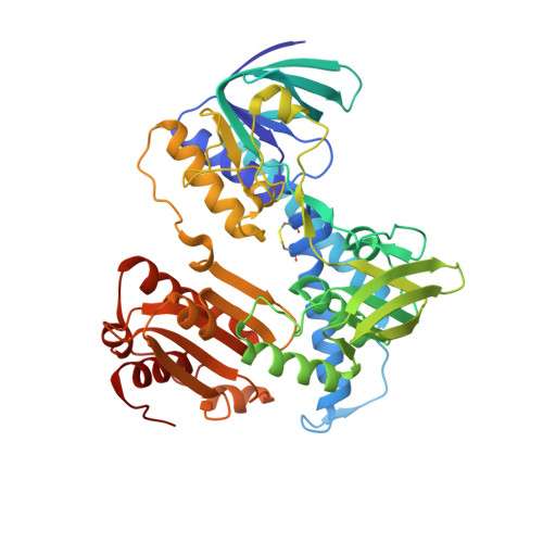
7KMY

Structure of Mtb Lpd bound to 010705


Experimental Data Snapshot

  • Method: X-RAY DIFFRACTION
  • Resolution: 2.21 Å
  • R-Value Free: 0.218 
  • R-Value Work: 0.181 
  • R-Value Observed: 0.183 

Starting Model: experimental
View more details

wwPDB Validation   3D Report Full Report


Ligand Structure Quality Assessment 


This is version 1.1 of the entry. See complete history


Literature

Whole Cell Active Inhibitors of Mycobacterial Lipoamide Dehydrogenase Afford Selectivity over the Human Enzyme through Tight Binding Interactions.

Ginn, J.Jiang, X.Sun, S.Michino, M.Huggins, D.J.Mbambo, Z.Jansen, R.Rhee, K.Y.Arango, N.Lima, C.D.Liverton, N.Imaeda, T.Okamoto, R.Kuroita, T.Aso, K.Stamford, A.Foley, M.Meinke, P.T.Nathan, C.Bryk, R.

(2021) ACS Infect Dis 7: 435-444

  • DOI: https://doi.org/10.1021/acsinfecdis.0c00788
  • Primary Citation of Related Structures:  
    7KMY

  • PubMed Abstract: 

    Tuberculosis remains a leading cause of death from a single bacterial infection worldwide. Efforts to develop new treatment options call for expansion into an unexplored target space to expand the drug pipeline and bypass resistance to current antibiotics. Lipoamide dehydrogenase is a metabolic and antioxidant enzyme critical for mycobacterial growth and survival in mice. Sulfonamide analogs were previously identified as potent and selective inhibitors of mycobacterial lipoamide dehydrogenase in vitro but lacked activity against whole mycobacteria. Here we present the development of analogs with improved permeability, potency, and selectivity, which inhibit the growth of Mycobacterium tuberculosis in axenic culture on carbohydrates and within mouse primary macrophages. They increase intrabacterial pyruvate levels, supporting their on-target activity within mycobacteria. Distinct modalities of binding between the mycobacterial and human enzymes contribute to improved potency and hence selectivity through induced-fit tight binding interactions within the mycobacterial but not human enzyme, as indicated by kinetic analysis and crystallography.


  • Organizational Affiliation

    Tri-Institutional Therapeutics Discovery Institute, New York, New York 10065, United States.


Macromolecules
Find similar proteins by:  (by identity cutoff)  |  3D Structure
Entity ID: 1
MoleculeChains Sequence LengthOrganismDetailsImage
Dihydrolipoyl dehydrogenase466Mycobacterium tuberculosisMutation(s): 0 
Gene Names: lpdClpdRv0462MTV038.06
EC: 1.8.1.4
UniProt
Find proteins for P9WHH9 (Mycobacterium tuberculosis (strain ATCC 25618 / H37Rv))
Explore P9WHH9 
Go to UniProtKB:  P9WHH9
Entity Groups  
Sequence Clusters30% Identity50% Identity70% Identity90% Identity95% Identity100% Identity
UniProt GroupP9WHH9
Sequence Annotations
Expand
  • Reference Sequence
Small Molecules
Ligands 3 Unique
IDChains Name / Formula / InChI Key2D Diagram3D Interactions
FAD
Query on FAD

Download Ideal Coordinates CCD File 
GB [auth N]
HA [auth D]
I [auth A]
LA [auth I]
Q [auth B]
GB [auth N],
HA [auth D],
I [auth A],
LA [auth I],
Q [auth B],
QA [auth J],
Y [auth C],
ZA [auth M]
FLAVIN-ADENINE DINUCLEOTIDE
C27 H33 N9 O15 P2
VWWQXMAJTJZDQX-UYBVJOGSSA-N
WPM (Subject of Investigation/LOI)
Query on WPM

Download Ideal Coordinates CCD File 
AB [auth M]
BB [auth M]
IA [auth D]
J [auth A]
K [auth A]
AB [auth M],
BB [auth M],
IA [auth D],
J [auth A],
K [auth A],
RA [auth J],
SA [auth J],
Z [auth C]
N~2~-methyl-N~2~-[(5-methyl-1H-indazol-7-yl)sulfonyl]-N-(1-methyl-2-oxo-1,2-dihydropyridin-4-yl)glycinamide
C17 H19 N5 O4 S
PMYKBSXBMRYDGS-UHFFFAOYSA-N
GOL
Query on GOL

Download Ideal Coordinates CCD File 
AA [auth C]
BA [auth C]
CA [auth C]
CB [auth M]
DA [auth C]
AA [auth C],
BA [auth C],
CA [auth C],
CB [auth M],
DA [auth C],
DB [auth M],
EA [auth C],
EB [auth M],
FA [auth C],
FB [auth M],
GA [auth C],
HB [auth N],
IB [auth N],
JA [auth D],
KA [auth D],
L [auth A],
M [auth A],
MA [auth I],
N [auth A],
NA [auth I],
O [auth A],
OA [auth I],
P [auth A],
PA [auth I],
R [auth B],
S [auth B],
T [auth B],
TA [auth J],
U [auth B],
UA [auth J],
V [auth B],
VA [auth J],
W [auth B],
WA [auth J],
X [auth B],
XA [auth J],
YA [auth J]
GLYCEROL
C3 H8 O3
PEDCQBHIVMGVHV-UHFFFAOYSA-N
Experimental Data & Validation

Experimental Data

  • Method: X-RAY DIFFRACTION
  • Resolution: 2.21 Å
  • R-Value Free: 0.218 
  • R-Value Work: 0.181 
  • R-Value Observed: 0.183 
  • Space Group: P 21 21 21
Unit Cell:
Length ( Å )Angle ( ˚ )
a = 107.506α = 90
b = 178.524β = 90
c = 226.445γ = 90
Software Package:
Software NamePurpose
PHENIXrefinement
PDB_EXTRACTdata extraction
HKL-2000data reduction
SCALEPACKdata scaling
PHENIXphasing

Structure Validation

View Full Validation Report


Ligand Structure Quality Assessment 


Entry History & Funding Information

Deposition Data

  • Released Date: 2021-01-27 
  • Deposition Author(s): Lima, C.D.

Funding OrganizationLocationGrant Number
National Institutes of Health/National Institute of General Medical Sciences (NIH/NIGMS)United StatesGM118080
National Institutes of Health/National Cancer Institute (NIH/NCI)United StatesP30CA008748
Howard Hughes Medical Institute (HHMI)United States--

Revision History  (Full details and data files)

  • Version 1.0: 2021-01-27
    Type: Initial release
  • Version 1.1: 2023-10-18
    Changes: Data collection, Database references, Refinement description