7KDU

Ricin bound to VHH antibody V5E4

  • Classification: TOXIN
  • Organism(s): Ricinus communis, Vicugna pacos
  • Expression System: Escherichia coli
  • Mutation(s): No 

  • Deposited: 2020-10-09 Released: 2021-08-04 
  • Deposition Author(s): Rudolph, M.J.
  • Funding Organization(s): National Institutes of Health/National Institute Of Allergy and Infectious Diseases (NIH/NIAID)

Experimental Data Snapshot

  • Method: X-RAY DIFFRACTION
  • Resolution: 2.81 Å
  • R-Value Free: 0.294 
  • R-Value Work: 0.237 
  • R-Value Observed: 0.240 

wwPDB Validation   3D Report Full Report


This is version 1.1 of the entry. See complete history


Literature

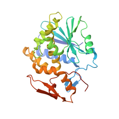
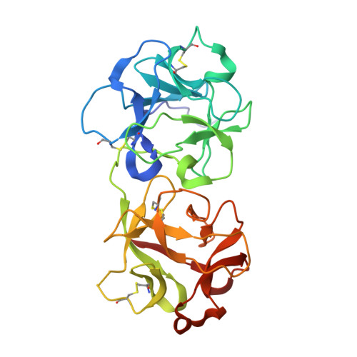
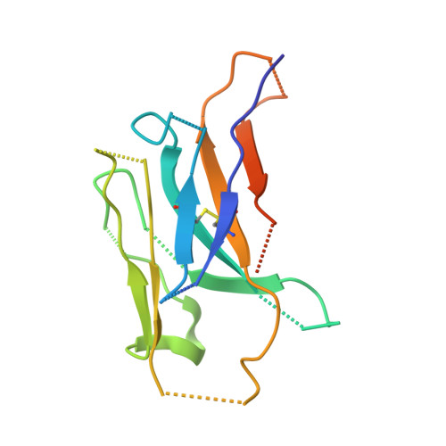
Structural Analysis of Toxin-Neutralizing, Single-Domain Antibodies that Bridge Ricin's A-B Subunit Interface.

Rudolph, M.J.Poon, A.Y.Kavaliauskiene, S.Myrann, A.G.Reynolds-Peterson, C.Davis, S.A.Sandvig, K.Vance, D.J.Mantis, N.J.

(2021) J Mol Biol 433: 167086-167086

  • DOI: https://doi.org/10.1016/j.jmb.2021.167086
  • Primary Citation of Related Structures:  
    7KBI, 7KBK, 7KC9, 7KD0, 7KD2, 7KDM, 7KDU

  • PubMed Abstract: 

    Ricin toxin kills mammalian cells with notorious efficiency. The toxin's B subunit (RTB) is a Gal/GalNAc-specific lectin that attaches to cell surfaces and promotes retrograde transport of ricin's A subunit (RTA) to the trans Golgi network (TGN) and endoplasmic reticulum (ER). RTA is liberated from RTB in the ER and translocated into the cell cytoplasm, where it functions as a ribosome-inactivating protein. While antibodies against ricin's individual subunits have been reported, we now describe seven alpaca-derived, single-domain antibodies (V H Hs) that span the RTA-RTB interface, including four Tier 1 V H Hs with IC 50 values <1 nM. Crystal structures of each V H H bound to native ricin holotoxin revealed three different binding modes, based on contact with RTA's F-G loop (mode 1), RTB's subdomain 2γ (mode 2) or both (mode 3). V H Hs in modes 2 and 3 were highly effective at blocking ricin attachment to HeLa cells and immobilized asialofetuin, due to framework residues (FR3) that occupied the 2γ Gal/GalNAc-binding pocket and mimic ligand. The four Tier 1 V H Hs also interfered with intracellular functions of RTB, as they neutralized ricin in a post-attachment cytotoxicity assay (e.g., the toxin was bound to cell surfaces before antibody addition) and reduced the efficiency of toxin transport to the TGN. We conclude that the RTA-RTB interface is a target of potent toxin-neutralizing antibodies that interfere with both extracellular and intracellular events in ricin's cytotoxic pathway.


  • Organizational Affiliation

    New York Structural Biology Center, New York, NY, USA. Electronic address: mrudolph@nysbc.org.


Macromolecules
Find similar proteins by:  (by identity cutoff)  |  3D Structure
Entity ID: 1
MoleculeChains Sequence LengthOrganismDetailsImage
Ricin chain A267Ricinus communisMutation(s): 0 
EC: 3.2.2.22
UniProt
Find proteins for P02879 (Ricinus communis)
Explore P02879 
Go to UniProtKB:  P02879
Entity Groups  
Sequence Clusters30% Identity50% Identity70% Identity90% Identity95% Identity100% Identity
UniProt GroupP02879
Sequence Annotations
Expand
  • Reference Sequence
Find similar proteins by:  (by identity cutoff)  |  3D Structure
Entity ID: 2
MoleculeChains Sequence LengthOrganismDetailsImage
Ricin chain B262Ricinus communisMutation(s): 0 
EC: 3.2.2.22
UniProt
Find proteins for P02879 (Ricinus communis)
Explore P02879 
Go to UniProtKB:  P02879
Entity Groups  
Sequence Clusters30% Identity50% Identity70% Identity90% Identity95% Identity100% Identity
UniProt GroupP02879
Sequence Annotations
Expand
  • Reference Sequence
Find similar proteins by:  (by identity cutoff)  |  3D Structure
Entity ID: 3
MoleculeChains Sequence LengthOrganismDetailsImage
VHH antibody V5E4133Vicugna pacosMutation(s): 0 
Entity Groups  
Sequence Clusters30% Identity50% Identity70% Identity90% Identity95% Identity100% Identity
Sequence Annotations
Expand
  • Reference Sequence
Oligosaccharides

Help

Entity ID: 4
MoleculeChains Length2D Diagram Glycosylation3D Interactions
alpha-L-fucopyranose-(1-3)-2-acetamido-2-deoxy-beta-D-glucopyranose
D
2N-Glycosylation
Glycosylation Resources
GlyTouCan:  G80587NA
GlyCosmos:  G80587NA
GlyGen:  G80587NA
Entity ID: 5
MoleculeChains Length2D Diagram Glycosylation3D Interactions
2-acetamido-2-deoxy-beta-D-glucopyranose-(1-4)-2-acetamido-2-deoxy-beta-D-glucopyranose
E, F
2N-Glycosylation
Glycosylation Resources
GlyTouCan:  G42666HT
GlyCosmos:  G42666HT
GlyGen:  G42666HT
Experimental Data & Validation

Experimental Data

  • Method: X-RAY DIFFRACTION
  • Resolution: 2.81 Å
  • R-Value Free: 0.294 
  • R-Value Work: 0.237 
  • R-Value Observed: 0.240 
  • Space Group: P 1 21 1
Unit Cell:
Length ( Å )Angle ( ˚ )
a = 45.281α = 90
b = 76.494β = 90.92
c = 101.44γ = 90
Software Package:
Software NamePurpose
PHENIXrefinement
SCALEPACKdata scaling
PDB_EXTRACTdata extraction
DENZOdata reduction
PHASERphasing

Structure Validation

View Full Validation Report


Entry History & Funding Information

Deposition Data


Funding OrganizationLocationGrant Number
National Institutes of Health/National Institute Of Allergy and Infectious Diseases (NIH/NIAID)United StatesHHSN272201400021C

Revision History  (Full details and data files)

  • Version 1.0: 2021-08-04
    Type: Initial release
  • Version 1.1: 2023-10-18
    Changes: Data collection, Database references, Refinement description