Structures and kinetics of Thermotoga maritima MetY reveal new insights into the predominant sulfurylation enzyme of bacterial methionine biosynthesis.
Brewster, J.L., Pachl, P., McKellar, J.L.O., Selmer, M., Squire, C.J., Patrick, W.M.(2021) J Biological Chem 296: 100797-100797
- PubMed: 34019879 Search on PubMedSearch on PubMed Central
- DOI: https://doi.org/10.1016/j.jbc.2021.100797
- Primary Citation Related Structures:
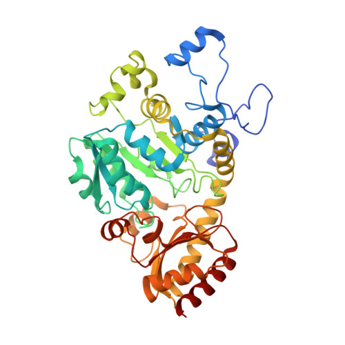
7KB0, 7KB1 - PubMed Abstract:
Bacterial methionine biosynthesis can take place by either the trans-sulfurylation route or direct sulfurylation. The enzymes responsible for trans-sulfurylation have been characterized extensively because they occur in model organisms such as Escherichia coli. However, direct sulfurylation is actually the predominant route for methionine biosynthesis across the phylogenetic tree. In this pathway, most bacteria use an O-acetylhomoserine aminocarboxypropyltransferase (MetY) to catalyze the formation of homocysteine from O-acetylhomoserine and bisulfide. Despite the widespread distribution of MetY, this pyridoxal 5'-phosphate-dependent enzyme remains comparatively understudied. To address this knowledge gap, we have characterized the MetY from Thermotoga maritima (TmMetY). At its optimal temperature of 70 °C, TmMetY has a turnover number (apparent k cat = 900 s -1 ) that is 10- to 700-fold higher than the three other MetY enzymes for which data are available. We also present crystal structures of TmMetY in the internal aldimine form and, fortuitously, with a β,γ-unsaturated ketimine reaction intermediate. This intermediate is identical to that found in the catalytic cycle of cystathionine γ-synthase (MetB), which is a homologous enzyme from the trans-sulfurylation pathway. By comparing the TmMetY and MetB structures, we have identified Arg270 as a critical determinant of specificity. It helps to wall off the active site of TmMetY, disfavoring the binding of the first MetB substrate, O-succinylhomoserine. It also ensures a strict specificity for bisulfide as the second substrate of MetY by occluding the larger MetB substrate, cysteine. Overall, this work illuminates the subtle structural mechanisms by which homologous pyridoxal 5'-phosphate-dependent enzymes can effect different catalytic, and therefore metabolic, outcomes.
- Department of Biochemistry, University of Otago, Dunedin, New Zealand.
Organizational Affiliation: