7K99

Crystal Structure of P. aeruginosa LpxC with N-Hydroxyformamide inhibitor 19


Experimental Data Snapshot

  • Method: X-RAY DIFFRACTION
  • Resolution: 1.90 Å
  • R-Value Free: 0.224 
  • R-Value Work: 0.166 
  • R-Value Observed: 0.169 

wwPDB Validation   3D Report Full Report


Ligand Structure Quality Assessment 


This is version 1.1 of the entry. See complete history


Literature

N-Hydroxyformamide LpxC inhibitors, their in vivo efficacy in a mouse Escherichia coli infection model, and their safety in a rat hemodynamic assay.

Furuya, T.Shapiro, A.B.Comita-Prevoir, J.Kuenstner, E.J.Zhang, J.Ribe, S.D.Chen, A.Hines, D.Moussa, S.H.Carter, N.M.Sylvester, M.A.Romero, J.A.C.Vega, C.V.Sacco, M.D.Chen, Y.O'Donnell, J.P.Durand-Reville, T.F.Miller, A.A.Tommasi, R.A.

(2020) Bioorg Med Chem 28: 115826-115826

  • DOI: https://doi.org/10.1016/j.bmc.2020.115826
  • Primary Citation of Related Structures:  
    7K99, 7K9A

  • PubMed Abstract: 

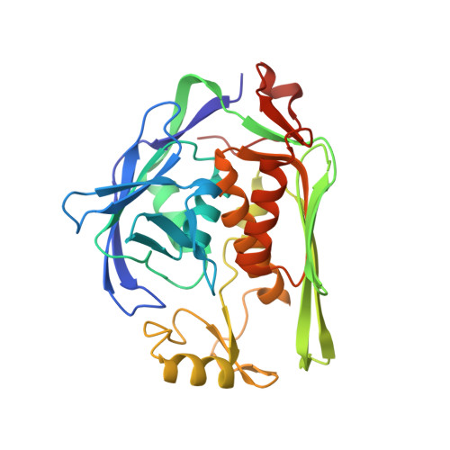
    UDP-3-O-(R-3-hydroxyacyl)-N-acetylglucosamine deacetylase (LpxC), the zinc metalloenzyme catalyzing the first committed step of lipid A biosynthesis in Gram-negative bacteria, has been a target for antibacterial drug discovery for many years. All inhibitor chemotypes reaching an advanced preclinical stage and clinical phase 1 have contained terminal hydroxamic acid, and none have been successfully advanced due, in part, to safety concerns, including hemodynamic effects. We hypothesized that the safety of LpxC inhibitors could be improved by replacing the terminal hydroxamic acid with a different zinc-binding group. After choosing an N-hydroxyformamide zinc-binding group, we investigated the structure-activity relationship of each part of the inhibitor scaffold with respect to Pseudomonas aeruginosa and Escherichia coli LpxC binding affinity, in vitro antibacterial potency and pharmacological properties. We identified a novel, potency-enhancing hydrophobic binding interaction for an LpxC inhibitor. We demonstrated in vivo efficacy of one compound in a neutropenic mouse E. coli infection model. Another compound was tested in a rat hemodynamic assay and was found to have a hypotensive effect. This result demonstrated that replacing the terminal hydroxamic acid with a different zinc-binding group was insufficient to avoid this previously recognized safety issue with LpxC inhibitors.


  • Organizational Affiliation

    Entasis Therapeutics, 35 Gatehouse Dr., Waltham, MA 02451, USA. Electronic address: takeru.furuya@astellas.com.


Macromolecules
Find similar proteins by:  (by identity cutoff)  |  3D Structure
Entity ID: 1
MoleculeChains Sequence LengthOrganismDetailsImage
UDP-3-O-acyl-N-acetylglucosamine deacetylaseA,
B [auth C]
303Pseudomonas aeruginosa PAO1Mutation(s): 0 
Gene Names: lpxCenvAPA4406
EC: 3.5.1.108
UniProt
Find proteins for P47205 (Pseudomonas aeruginosa (strain ATCC 15692 / DSM 22644 / CIP 104116 / JCM 14847 / LMG 12228 / 1C / PRS 101 / PAO1))
Explore P47205 
Go to UniProtKB:  P47205
Entity Groups  
Sequence Clusters30% Identity50% Identity70% Identity90% Identity95% Identity100% Identity
UniProt GroupP47205
Sequence Annotations
Expand
  • Reference Sequence
Small Molecules
Ligands 4 Unique
IDChains Name / Formula / InChI Key2D Diagram3D Interactions
W4M (Subject of Investigation/LOI)
Query on W4M

Download Ideal Coordinates CCD File 
D [auth A],
M [auth C]
(hydroxy{(1S)-1-(methylsulfanyl)-2-[5-({4-[(morpholin-4-yl)methyl]phenyl}ethynyl)-1H-benzotriazol-1-yl]ethyl}amino)methanol
C23 H27 N5 O3 S
AMIXUMKMQGGIQQ-QHCPKHFHSA-N
SO4
Query on SO4

Download Ideal Coordinates CCD File 
E [auth A]
F [auth A]
G [auth A]
H [auth A]
N [auth C]
E [auth A],
F [auth A],
G [auth A],
H [auth A],
N [auth C],
O [auth C],
P [auth C],
Q [auth C]
SULFATE ION
O4 S
QAOWNCQODCNURD-UHFFFAOYSA-L
GOL
Query on GOL

Download Ideal Coordinates CCD File 
C [auth A],
I [auth A],
J [auth A],
L [auth C],
R [auth C]
GLYCEROL
C3 H8 O3
PEDCQBHIVMGVHV-UHFFFAOYSA-N
ZN
Query on ZN

Download Ideal Coordinates CCD File 
K [auth A],
S [auth C]
ZINC ION
Zn
PTFCDOFLOPIGGS-UHFFFAOYSA-N
Experimental Data & Validation

Experimental Data

  • Method: X-RAY DIFFRACTION
  • Resolution: 1.90 Å
  • R-Value Free: 0.224 
  • R-Value Work: 0.166 
  • R-Value Observed: 0.169 
  • Space Group: P 1 21 1
Unit Cell:
Length ( Å )Angle ( ˚ )
a = 35.763α = 90
b = 158.047β = 101.9
c = 49.072γ = 90
Software Package:
Software NamePurpose
HKL-2000data scaling
REFMACrefinement
PDB_EXTRACTdata extraction
HKL-3000data reduction
MOLREPphasing

Structure Validation

View Full Validation Report


Ligand Structure Quality Assessment 


Entry History 

Deposition Data

Revision History  (Full details and data files)

  • Version 1.0: 2020-11-25
    Type: Initial release
  • Version 1.1: 2023-10-18
    Changes: Data collection, Database references, Refinement description