7K66

Structure of Blood Coagulation Factor VIII in Complex with an Anti-C1 Domain Pathogenic Antibody Inhibitor


Experimental Data Snapshot

  • Method: X-RAY DIFFRACTION
  • Resolution: 3.92 Å
  • R-Value Free: 0.322 
  • R-Value Work: 0.226 
  • R-Value Observed: 0.234 

wwPDB Validation   3D Report Full Report


Ligand Structure Quality Assessment 


This is version 1.3 of the entry. See complete history


Literature

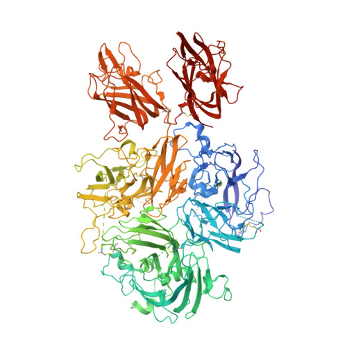
Structure of blood coagulation factor VIII in complex with an anti-C1 domain pathogenic antibody inhibitor.

Gish, J.S.Jarvis, L.Childers, K.C.Peters, S.C.Garrels, C.S.Smith, I.W.Spencer, H.T.Doering, C.B.Lollar, P.Spiegel, P.C.

(2021) Blood 137: 2981-2986

  • DOI: https://doi.org/10.1182/blood.2020008940
  • Primary Citation of Related Structures:  
    7K66

  • PubMed Abstract: 

    Antibody inhibitor development in hemophilia A represents the most significant complication resulting from factor VIII (fVIII) replacement therapy. Recent studies have demonstrated that epitopes present in the C1 domain contribute to a pathogenic inhibitor response. In this study, we report the structure of a group A anti-C1 domain inhibitor, termed 2A9, in complex with a B domain-deleted, bioengineered fVIII construct (ET3i). The 2A9 epitope forms direct contacts to the C1 domain at 3 different surface loops consisting of Lys2065-Trp2070, Arg2150-Tyr2156, and Lys2110-Trp2112. Additional contacts are observed between 2A9 and the A3 domain, including the Phe1743-Tyr1748 loop and the N-linked glycosylation at Asn1810. Most of the C1 domain loops in the 2A9 epitope also represent a putative interface between fVIII and von Willebrand factor. Lastly, the C2 domain in the ET3i:2A9 complex adopts a large, novel conformational change, translocating outward from the structure of fVIII by 20 Å. This study reports the first structure of an anti-C1 domain antibody inhibitor and the first fVIII:inhibitor complex with a therapeutically active fVIII construct. Further structural understanding of fVIII immunogenicity may result in the development of more effective and safe fVIII replacement therapies.


  • Organizational Affiliation

    Department of Chemistry, Western Washington University, Bellingham, WA; and.


Macromolecules
Find similar proteins by:  (by identity cutoff)  |  3D Structure
Entity ID: 1
MoleculeChains Sequence LengthOrganismDetailsImage
Coagulation factor VIII,Coagulation factor VIII,Coagulation factor VIII,Coagulation factor VIII1,467Sus scrofaHomo sapiensMutation(s): 0 
Gene Names: F8CF8F8C
Membrane Entity: Yes 
UniProt & NIH Common Fund Data Resources
Find proteins for P12263 (Sus scrofa)
Explore P12263 
Go to UniProtKB:  P12263
Find proteins for P00451 (Homo sapiens)
Explore P00451 
Go to UniProtKB:  P00451
PHAROS:  P00451
GTEx:  ENSG00000185010 
Entity Groups  
Sequence Clusters30% Identity50% Identity70% Identity90% Identity95% Identity100% Identity
UniProt GroupsP12263P00451
Sequence Annotations
Expand
  • Reference Sequence
Find similar proteins by:  (by identity cutoff)  |  3D Structure
Entity ID: 2
MoleculeChains Sequence LengthOrganismDetailsImage
2A9 heavy chain223Mus musculusMutation(s): 0 
Entity Groups  
Sequence Clusters30% Identity50% Identity70% Identity90% Identity95% Identity100% Identity
Sequence Annotations
Expand
  • Reference Sequence
Find similar proteins by:  (by identity cutoff)  |  3D Structure
Entity ID: 3
MoleculeChains Sequence LengthOrganismDetailsImage
2A9 light chain213Mus musculusMutation(s): 0 
Entity Groups  
Sequence Clusters30% Identity50% Identity70% Identity90% Identity95% Identity100% Identity
Sequence Annotations
Expand
  • Reference Sequence
Oligosaccharides

Help

Entity ID: 4
MoleculeChains Length2D Diagram Glycosylation3D Interactions
alpha-D-mannopyranose-(1-3)-[alpha-D-mannopyranose-(1-6)]beta-D-mannopyranose-(1-4)-2-acetamido-2-deoxy-beta-D-glucopyranose-(1-4)-2-acetamido-2-deoxy-beta-D-glucopyranoseD [auth H]5N-Glycosylation
Glycosylation Resources
GlyTouCan:  G22768VO
GlyCosmos:  G22768VO
GlyGen:  G22768VO
Entity ID: 5
MoleculeChains Length2D Diagram Glycosylation3D Interactions
2-acetamido-2-deoxy-beta-D-glucopyranose-(1-4)-2-acetamido-2-deoxy-beta-D-glucopyranoseE [auth D]2N-Glycosylation
Glycosylation Resources
GlyTouCan:  G42666HT
GlyCosmos:  G42666HT
GlyGen:  G42666HT
Experimental Data & Validation

Experimental Data

  • Method: X-RAY DIFFRACTION
  • Resolution: 3.92 Å
  • R-Value Free: 0.322 
  • R-Value Work: 0.226 
  • R-Value Observed: 0.234 
  • Space Group: P 41 21 2
Unit Cell:
Length ( Å )Angle ( ˚ )
a = 116.721α = 90
b = 116.721β = 90
c = 371.057γ = 90
Software Package:
Software NamePurpose
Adxvdata reduction
XDSdata reduction
PHASERphasing
Cootmodel building
PHENIXrefinement
Aimlessdata scaling

Structure Validation

View Full Validation Report


Ligand Structure Quality Assessment 


Entry History & Funding Information

Deposition Data


Funding OrganizationLocationGrant Number
National Science Foundation (NSF, United States)United StatesMRI 1429164
National Institutes of Health/National Heart, Lung, and Blood Institute (NIH/NHLBI)United StatesR15HL103518
National Institutes of Health/National Heart, Lung, and Blood Institute (NIH/NHLBI)United StatesU54HL141981
National Institutes of Health/National Heart, Lung, and Blood Institute (NIH/NHLBI)United StatesR44HL117511
National Institutes of Health/National Heart, Lung, and Blood Institute (NIH/NHLBI)United StatesR44HL110448
National Institutes of Health/National Heart, Lung, and Blood Institute (NIH/NHLBI)United StatesU54HL112309

Revision History  (Full details and data files)

  • Version 1.0: 2020-10-14
    Type: Initial release
  • Version 1.1: 2021-02-24
    Changes: Database references
  • Version 1.2: 2021-06-09
    Changes: Database references
  • Version 1.3: 2023-10-18
    Changes: Data collection, Database references, Refinement description